. 2024 Jun;29(6):1869-1881.
doi: 10.1038/s41380-024-02442-7. Epub 2024 Feb 9.

Connectome architecture shapes large-scale cortical alterations in schizophrenia: a worldwide ENIGMA study

Foivos Georgiadis  1 Sara Larivière  2 David Glahn  3 L Elliot Hong  4 Peter Kochunov  4 Bryan Mowry  5 Carmel Loughland  6 Christos Pantelis  7 Frans A Henskens  8 Melissa J Green  9 Murray J Cairns  10 Patricia T Michie  11 Paul E Rasser  12 Stanley Catts  13 Paul Tooney  10   14 Rodney J Scott  10 Ulrich Schall  14 Vaughan Carr  15 Yann Quidé  9 Axel Krug  16 Frederike Stein  17 Igor Nenadić  18 Katharina Brosch  17 Tilo Kircher  17 Raquel Gur  19 Ruben Gur  19 Theodore D Satterthwaite  19 Andriana Karuk  20 Edith Pomarol-Clotet  20 Joaquim Radua  21 Paola Fuentes-Claramonte  20 Raymond Salvador  20 Gianfranco Spalletta  22 Aristotle Voineskos  10 Kang Sim  23 Benedicto Crespo-Facorro  24 Diana Tordesillas Gutiérrez  25 Stefan Ehrlich  26 Nicolas Crossley  27 Dominik Grotegerd  28 Jonathan Repple  28 Rebekka Lencer  28 Udo Dannlowski  28 Vince Calhoun  29 Kelly Rootes-Murdy  29 Caroline Demro  30   31 Ian S Ramsay  32 Scott R Sponheim  31   32 Andre Schmidt  33 Stefan Borgwardt  34 Alexander Tomyshev  35 Irina Lebedeva  35 Cyril Höschl  36 Filip Spaniel  36 Adrian Preda  37 Dana Nguyen  38 Anne Uhlmann  39 Dan J Stein  40 Fleur Howells  40 Henk S Temmingh  40 Ana M Diaz Zuluaga  41 Carlos López Jaramillo  41 Felice Iasevoli  42 Ellen Ji  43 Stephanie Homan  43 Wolfgang Omlor  43 Philipp Homan  43 Stefan Kaiser  44 Erich Seifritz  43 Bratislav Misic  2 Sofie L Valk  45   46 Paul Thompson  47 Theo G M van Erp  37 Jessica A Turner  48 ENIGMA Schizophrenia ConsortiumBoris Bernhardt #  2 Matthias Kirschner #  49   50
Affiliations

Connectome architecture shapes large-scale cortical alterations in schizophrenia: a worldwide ENIGMA study

Foivos Georgiadis et al. Mol Psychiatry. 2024 Jun.

Abstract

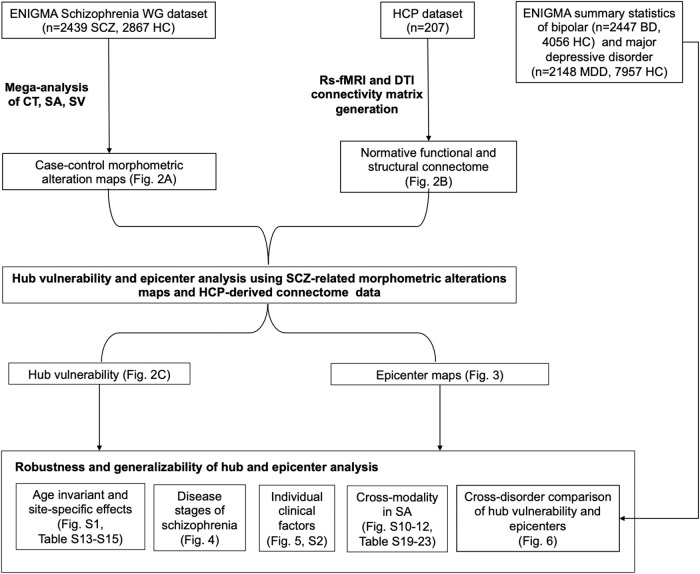
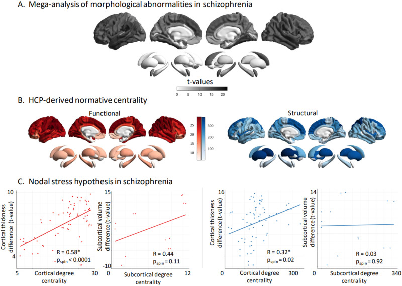
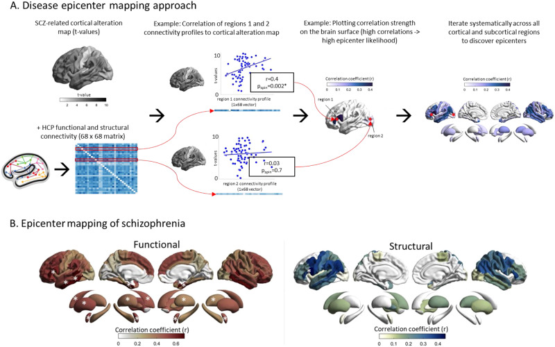
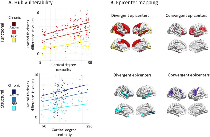
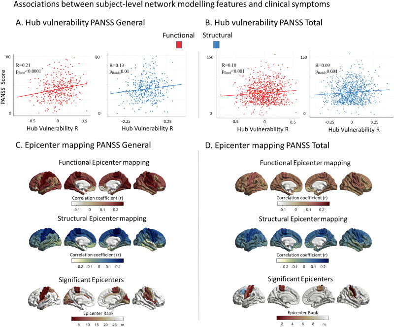
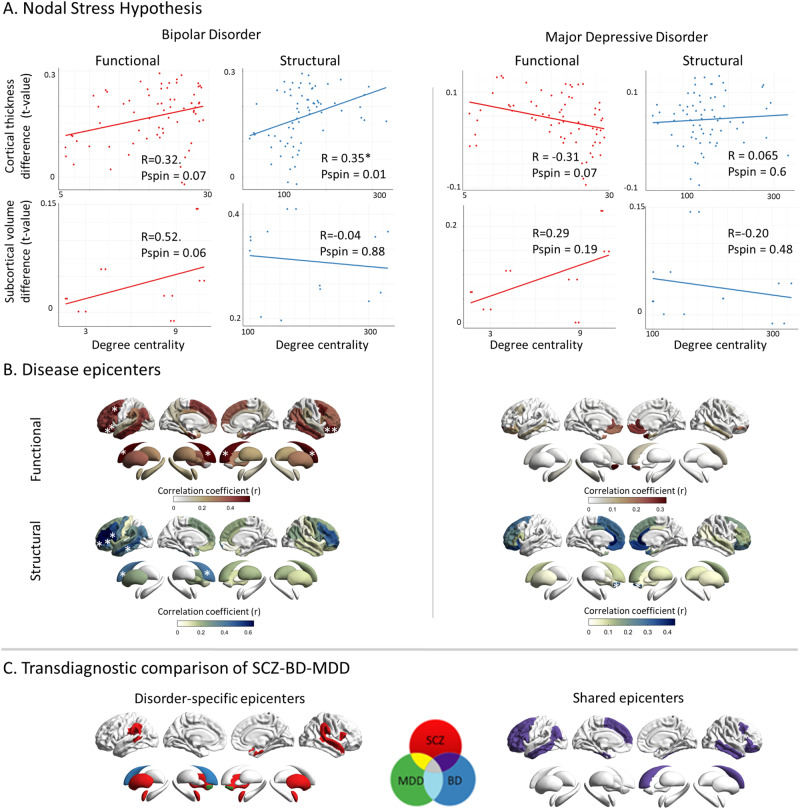
Schizophrenia is a prototypical network disorder with widespread brain-morphological alterations, yet it remains unclear whether these distributed alterations robustly reflect the underlying network layout. We tested whether large-scale structural alterations in schizophrenia relate to normative structural and functional connectome architecture, and systematically evaluated robustness and generalizability of these network-level alterations. Leveraging anatomical MRI scans from 2439 adults with schizophrenia and 2867 healthy controls from 26 ENIGMA sites and normative data from the Human Connectome Project (n = 207), we evaluated structural alterations of schizophrenia against two network susceptibility models: (i) hub vulnerability, which examines associations between regional network centrality and magnitude of disease-related alterations; (ii) epicenter mapping, which identifies regions whose typical connectivity profile most closely resembles the disease-related morphological alterations. To assess generalizability and specificity, we contextualized the influence of site, disease stages, and individual clinical factors and compared network associations of schizophrenia with that found in affective disorders. Our findings show schizophrenia-related cortical thinning is spatially associated with functional and structural hubs, suggesting that highly interconnected regions are more vulnerable to morphological alterations. Predominantly temporo-paralimbic and frontal regions emerged as epicenters with connectivity profiles linked to schizophrenia's alteration patterns. Findings were robust across sites, disease stages, and related to individual symptoms. Moreover, transdiagnostic comparisons revealed overlapping epicenters in schizophrenia and bipolar, but not major depressive disorder, suggestive of a pathophysiological continuity within the schizophrenia-bipolar-spectrum. In sum, cortical alterations over the course of schizophrenia robustly follow brain network architecture, emphasizing marked hub susceptibility and temporo-frontal epicenters at both the level of the group and the individual. Subtle variations of epicenters across disease stages suggest interacting pathological processes, while associations with patient-specific symptoms support additional inter-individual variability of hub vulnerability and epicenters in schizophrenia. Our work outlines potential pathways to better understand macroscale structural alterations, and inter- individual variability in schizophrenia.


Conflict of interest statement

The authors declare no competing interests.

Figures

Fig. 1
Fig. 1. Flowchart of multi-source data integration and analysis steps.
BD bipolar disorder, HC healthy controls, HCP Human Connectome Project, MDD major depressive disorder; SA surface area, SCZ schizophrenia, SV subcortical volume; WG Working Group.
Fig. 2
Fig. 2. Hub vulnerability mapping.
A Unthresholded t-maps of cortical thickness and subcortical volume deficiencies in SCZ (n = 2439), compared to HCP (n = 2867). B Normative functional and structural network organization, derived from the HCP dataset, was used to identify hubs (i.e., regions with greater degree centrality). C Correlation of gray matter morphological alterations with node-level functional (left) and structural (right) maps of degree centrality. In SCZ, regions with high functional or structural centrality are significantly more likely to display morphological alterations in the cortex. Subcortical functional subcortico-cortical degree centrality showed a moderate non-significant correlation with morphometric alterations, while no correlation was observed for structural subcortico-cortical degree centrality. HCP healthy control participants, SCZ schizophrenia.
Fig. 3
Fig. 3. Disease epicenters.
A Disease epicenter mapping schematic for cortico-cortical connectivity: To discover epicenters of SCZ, the SCZ-related cortical alteration t-value vector (1 × 68) from our mega-analysis, was iteratively correlated with each region-specific cortico-cortical connectivity vector (1 × 68), derived from the HCP functional and structural connectivity matrices (68 × 68). In the example we showcase the procedure for two theoretical cortical regions with different magnitudes of correlations between each region’s cortico-cortical connectivity vector and the SCZ-specific t-value map. Regions whose connectivity vectors are highly correlated with SCZ-specific cortical alteration patterns (t-values) are likely epicenters of SCZ (Region 1). Regions whose connectivity vectors are weakly correlated with the SCZ-specific cortical alteration patterns (t-values) are unlikely epicenters of SCZ (Region 2). We systematically repeated this analysis across all cortical and subcortical regions for both functional and structural connectivity. The resulting correlation values of each region were plotted on the surface to generate epicenter maps that display the epicenter likelihood of each region. B Correlation coefficient maps depicting the strength of association between the normative region-based functional (left) and structural (right) connectivity and the SCZ-specific morphological alteration maps. Disease epicenters are regions that are more strongly connected to regions with more pronounced morphometric alterations - and, inversely, are more weakly connected with regions with less pronounced alterations. Asterisks denote the top five significant epicenters. Top-5 functional epicenters, cortical: entorhinal cortex (L + R), banks of superior temporal sulcus (L,), inferior temporal gyrus (L); subcortical: amygdala (L + R), putamen (L + R), caudate (L). Top-5 structural epicenters, cortical: pars opercularis of inferior frontal gyrus (L); subcortical: none.
Fig. 4
Fig. 4. Disease stage comparison of network modeling.
A Correlation of cortical thickness abnormality in SCZ to node- level functional (upper) and structural (lower) maps of degree centrality (all r > 0.23, pspin< 0.05). Color code indicates first- episode, early disease stage, and chronic disease stage from brighter to darker colors. B SCZ epicenter mapping (for details, see Fig. 2). Divergent (unique) epicenters are only significant in one of three disease stages with color indicating the corresponding disease stage. Convergent epicenters are significant in all three disease stages (pspin < 0.05). Subgroups of disease stages are defined by duration of illness: first-episode psychosis (<1 month); early disease stage (>1 month but <2 years); chronic disease stage (>20 years). FEP First episode psychosis.
Fig. 5
Fig. 5. Associations between subject-level network modeling and individual symptoms.
A Correlation between subject-level functional (red) and structural (blue) hub vulnerability of cortical alterations and PANSS general scores (rfunc = 0.21, pBonf < 0.0001, rstruc = 0.13, pBonf = 0.01). B Correlation between subject-level functional (red) and structural (blue) hub vulnerability of cortical alterations and PANSS total scores (rfunc = 0.10, pBonf = 0.001, rstruc = 0.09, pBonf = 0.004). C Correlations between subject-level functional (red) and structural (blue) epicenter likelihood and PANSS general scores. Significant associations were only found for functional epicenters spanning from sensory-motor areas to cingulate gyrus and insula (all r > .18, pBonf < 0.05). D Correlations between subject-level functional (red) and structural (blue) epicenter likelihood and PANSS total scores. Significant associations were found for functional epicenters in the bilateral somatosensory and motor cortices extending to supramarginal gyrus (in blue) as the only structural epicenter ((all r > 0.11, pBonf < 0.05).
Fig. 6
Fig. 6. Cross-disorder comparison of network modeling.
A Correlation of disorder-related gray matter alterations to node-level functional (left) and structural (right) maps of degree centrality in BD and MDD. In BD, cortical regions with high structural centrality are significantly more likely to display higher morphological alterations; this trend is also observed for functional centrality. No such relationship is observed in MDD. B Correlation coefficient maps depicting strength of association between the normative region-based functional (top) and structural (bottom) connectivity and the BD- (left) and MDD-specific (right) morphological alteration maps. Asterisks denote the significant epicenters. Functional BD epicenters: pars orbitalis of inferior frontal gyrus (L), lateral orbitofrontal cortex (L), caudal middle frontal gyrus (L), inferior and middle temporal gyrus (L) cortically and the caudate nucleus (L) in the subcortex (func: bilateral, struc: left). Structural BD epicenters: pars opercularis (L) and pars triangularis (L) of inferior frontal gyrus, rostral middle frontal gyrus (L), middle temporal gyrus (L) cortically and the caudate nucleus (L), subcortically. In MDD, no functional epicenters were detected. Structural MDD epicenters: nucleus accumbens (R). C Transdiagnostic comparison of epicenters between SCZ, BD, and MDD. Disorder- specific epicenters which are only significant in one disorder are displayed on the left side. Shared epicenters between at least two disorders are shown on the right side. Please note, that shared epicenters were only found for SCZ and BD but not MDD: Bipolar disorder; MDD: Major depressive disorder; SCZ: Schizophrenia.

Comment in

References

    1. Fornito A, Zalesky A, Breakspear M. The connectomics of brain disorders. Nat Rev Neurosci. 2015;16:159–72. 10.1038/nrn3901 - DOI - PubMed
    1. Hansen JY, Shafiei G, Vogel JW, Smart K, Bearden CE, Hoogman M, et al. Local molecular and global connectomic contributions to cross-disorder cortical abnormalities. Nat Commun. 2022;13:1–17. 10.1038/s41467-022-32420-y - DOI - PMC - PubMed
    1. Repple J, Gruber M, Mauritz M, de Lange SC, Winter NR, Opel N, et al. Shared and specific patterns of structural brain connectivity across affective and psychotic disorders. Biol Psychiatry. 2022;93:178–86. 10.1016/j.biopsych.2022.05.031 - DOI - PubMed
    1. van den Heuvel MP, Sporns O. A cross-disorder connectome landscape of brain dysconnectivity. Nat Rev Neurosci. 2019;20:435–46. 10.1038/s41583-019-0177-6 - DOI - PMC - PubMed
    1. van Erp TGM, Walton E, Hibar DP, Schmaal L, Jiang W, Glahn DC, et al. Cortical brain abnormalities in 4474 individuals with schizophrenia and 5098 control subjects via the Enhancing Neuro Imaging Genetics Through Meta Analysis (ENIGMA) Consortium. Biol Psychiatry. 2018;84:644–54. 10.1016/j.biopsych.2018.04.023 - DOI - PMC - PubMed