Clinical Trial
. 2024 Feb 9;14(1):3379.
doi: 10.1038/s41598-024-54041-9.

Immunological characterization of a long-lasting response in a patient with metastatic triple-negative breast cancer treated with PD-1 and LAG-3 blockade

Affiliations
Clinical Trial

Immunological characterization of a long-lasting response in a patient with metastatic triple-negative breast cancer treated with PD-1 and LAG-3 blockade

Licia Rivoltini et al. Sci Rep. .

Abstract

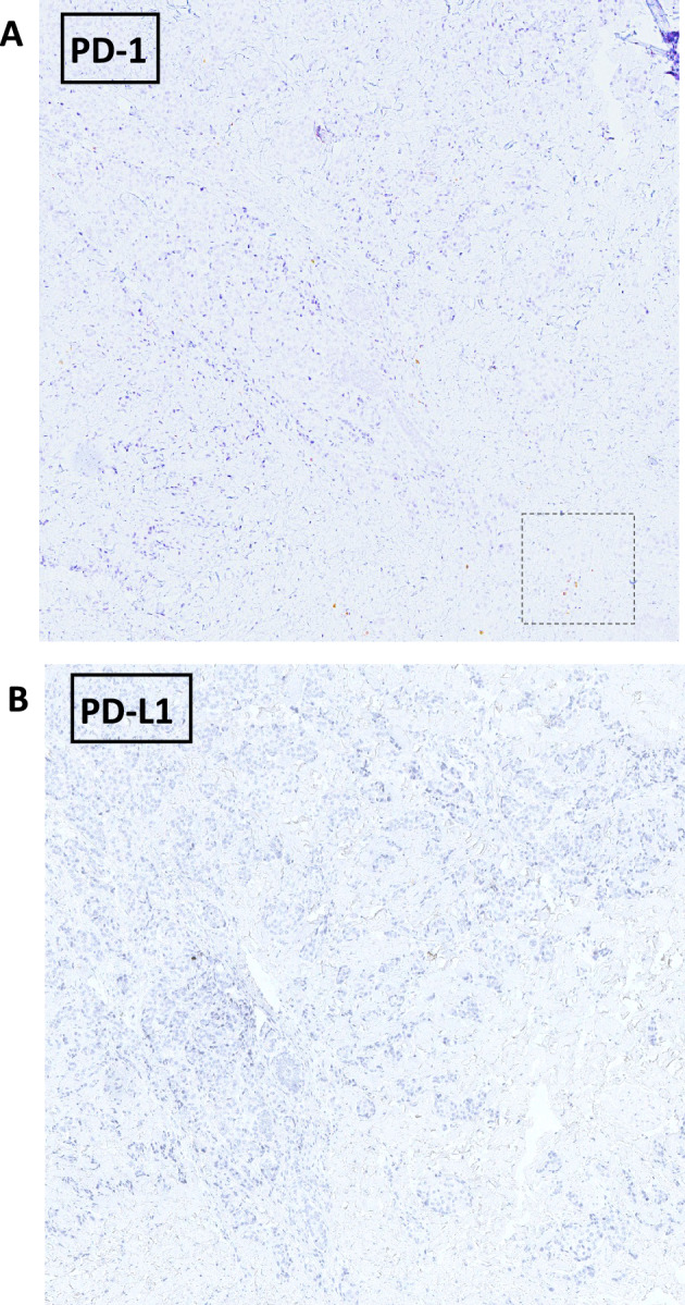
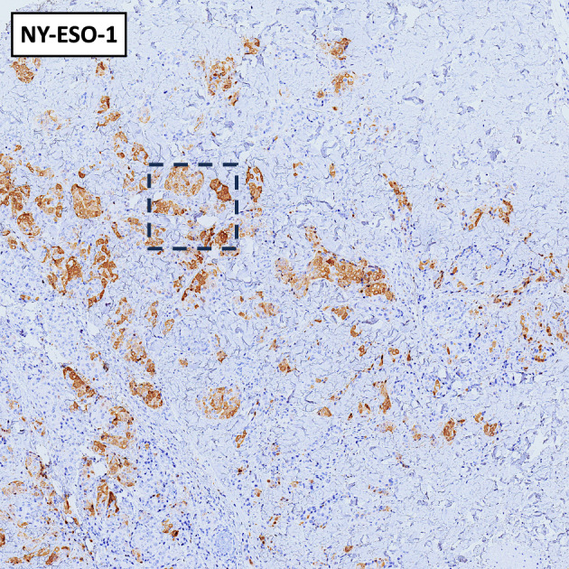
In patients with advanced triple-negative breast cancer (TNBC), translational research efforts are needed to improve the clinical efficacy of immunotherapy with checkpoint inhibitors. Here, we report on the immunological characterization of an exceptional, long-lasting, tumor complete response in a patient with metastatic TNBC treated with dual PD-1 and LAG-3 blockade within the phase I/II study CLAG525X2101C (NCT02460224) The pre-treatment tumor biopsy revealed the presence of a CD3+ and CD8+ cell infiltrate, with few PD1+ cells, rare CD4+ cells, and an absence of both NK cells and LAG3 expression. Conversely, tumor cells exhibited positive staining for the three primary LAG-3 ligands (HLA-DR, FGL-1, and galectin-3), while being negative for PD-L1. In peripheral blood, baseline expression of LAG-3 and PD-1 was observed in circulating immune cells. Following treatment initiation, there was a rapid increase in proliferating granzyme-B+ NK and T cells, including CD4+ T cells, alongside a reduction in myeloid-derived suppressor cells. The role of LAG-3 expression on circulating NK cells, as well as the expression of LAG-3 ligands on tumor cells and the early modulation of circulating cytotoxic CD4+ T cells warrant further investigation as exploitable predictive biomarkers for dual PD-1 and LAG-3 blockade.Trial registration: NCT02460224. Registered 02/06/2015.


Conflict of interest statement

The authors declare no competing interests.

Figures

Figure 1
Figure 1
Dynamics of response to dual PD-1/LAG-3 blockade and immune-molecular tumor characterization. (A) Shows the timeline of response dynamics together with the time of blood sampling for the immune monitoring. A rapid antitumor activity with a radiological partial response (as illustrated by the computed tomography images of a lymph node metastases in B) was achieved at week + 9 and was paralleled by the regression of most of the skin involvement. A complete clinical response of the extensive skin localization was observed at week + 18 (C). A complete radiological response (as per RECIST v.1.1) was achieved at week + 20. The immunohistochemical characterization of the tumor microenvironment showed a zonal CD3+ infiltrate mainly composed of CD8+ cells with rare CD4+ and rare PD-1+ cells (D, images at 20 × magnification). Tumor cells stained positive for the three main known LAG-3 ligands (HLA-DR, FGL-1 and galectin-3) and for the cancer testis antigen NY-ESO-1 (E, images at 20 × magnification).
Figure 2
Figure 2
Immunohistochemical staining at 4 × magnification. Immunohistochemical staining for PD-1 (A) and PD-L1 (B) at 4 × magnification. The dashed line rectangle indicates the area showed at 20 × magnification in Fig. 1D.
Figure 3
Figure 3
Immunohistochemical staining for NY-ESO-1 at 4 × magnification. The dashed line rectangle indicates the area showed at 20 × magnification in Fig. 1E.
Figure 4
Figure 4
Effects of PD-1/LAG-3 dual blockade on blood immune cell frequency and induction of a lymphocyte-mediated antitumor response soon after treatment initiation. Freshly thawed PBMCs were analysed by flow cytometry. (A) Percentage of LAG-3 (red) and PD-1 (blue) positive cells at the baseline in CD4+, CD8+, Treg (CD4+CD25+Foxp3+) and NK cells (CD16CD56hi and CD16+CD56dim) are reported in graph. (B) Frequency of lymphoid (CD3+, CD4+, CD8+, Treg, NK cells) and myeloid (CD14+ monocytes, M-MDSC and PMN-MDSC) populations at the baseline (pre) and after treatments (post 1,2, 7) are shown. (C) overlay dot plots show the increased proliferation of GrZB+ cells (rectangle gate) after the first treatment (red) compared to the baseline (green) in CD4+ and CD8+ T cells (upper row) and in NK CD56hi and NK CD56dim (lower row). The frequency of cells expressing both the intracellular markers, Ki67 and GrZB, are reported in the graph as percentages (red = first treatment, green = baseline). (D) The presence of NY-ESO-1 specific CD8+ T cells was evaluated comparing PBMCs at the baseline and after the first treatment using HLA-A*0201/NY-ESO-1 multimer staining. The percentage of CD8+ multimer positive cells was calculated in the CD8+CD19 gate and reported in the figure. (E) PMBCs were stained with a combination of mAbs to define naïve and memory T cell subsets. Histograms show the percentage of naïve (CD45RA+CCR7+, grey), effector memory (EM, CD45RACCR7, blue), central memory (CM, CD45RACCR7+, yellow) and terminally effector (TE, CD45RA+CCR7, red) cells in CD4+ and CD8+ lymphocytes at the baseline (pre), after the first treatment (post 1) and late in the treatment (post 48).

References

    1. Keenan TE, Tolaney SM. Role of immunotherapy in triple-negative breast cancer. J. Natl. Compr. Cancer Netw. 2020;18(4):479–489. doi: 10.6004/jnccn.2020.7554. - DOI - PubMed
    1. Fucà G, de Braud F, Di Nicola M. Immunotherapy-based combinations: an update. Curr. Opin. Oncol. 2018;30(5):345–351. doi: 10.1097/CCO.0000000000000466. - DOI - PubMed
    1. Maruhashi T, Sugiura D, Okazaki IM, Okazaki T. LAG-3: from molecular functions to clinical applications. J. Immunother. Cancer. 2020;8(2):e01001014. doi: 10.1136/jitc-2020-001014. - DOI - PMC - PubMed
    1. Hong DS, et al. Phase I/II study of LAG525 ± spartalizumab (PDR001) in patients (pts) with advanced malignancies. J. Clin. Oncol. 2018;36(15_Suppl):3012. doi: 10.1200/JCO.2018.36.15_suppl.3012. - DOI
    1. Pruneri G, Vingiani A, Bagnardi V, Rotmensz N, De Rose A, Palazzo A, et al. Clinical validity of tumor-infiltrating lymphocytes analysis in patients with triple-negative breast cancer. Ann. Oncol. 2016;27(2):249–256. doi: 10.1093/annonc/mdv571. - DOI - PubMed

Publication types

MeSH terms

Associated data