

Nerves and availability of mesodermal cells are essential for the function of the segment addition zone (SAZ) during segment regeneration in polychaete annelids
- PMID: 38336874
- PMCID: PMC11611952
- DOI: 10.1007/s00427-024-00713-5
Nerves and availability of mesodermal cells are essential for the function of the segment addition zone (SAZ) during segment regeneration in polychaete annelids
Abstract
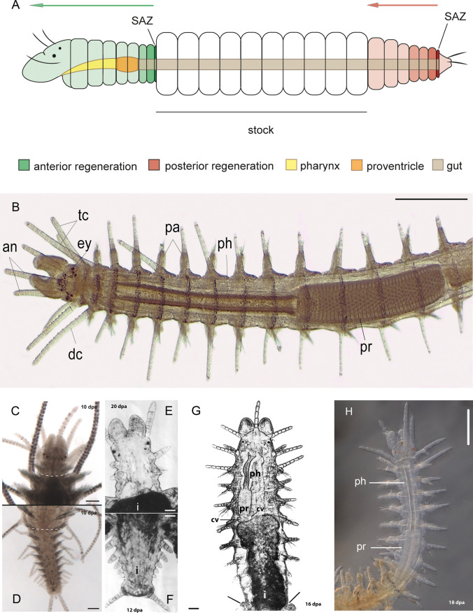
Most of annelids grow all over their asexual life through the continuous addition of segments from a special zone called "segment addition zone" (SAZ) adjacent to the posterior extremity called pygidium. Amputation of posterior segments leads to regeneration (posterior regeneration-PR) of the pygidium and a new SAZ, as well as new segments issued from this new SAZ. Amputation of anterior segments leads some species to regeneration (anterior regeneration-AR) of the prostomium and a SAZ which produces new segments postero-anteriorly as during PR. During the 1960s and 1970s decades, experimental methods on different species (Syllidae, Nereidae, Aricidae) showed that the function of SAZ depends on the presence and number of mesodermal regeneration cells. Selective destruction of mesodermal regeneration cells in AR had no effect on the regeneration of the prostomium, but as for PR, it inhibited segment regeneration. Thus, worms deprived of mesodermal regeneration cells are always able to regenerate the pygidium or the prostomium, but they are unable to regenerate segments, a result which indicates that the SAZ functions only if these regeneration cells are present during PR or AR. Additionally, during AR, nerve fibres regenerate from the cut nerve cord toward the newformed brain, a situation which deprives the SAZ of local regenerating nerve fibres and their secreted growth factors. In contrast, during PR, nerve fibres regenerate both during the entire regeneration phase and then in normal growth. This review summarizes the experimental evidence for mesoderm cell involvement in segment regeneration, and the differential impact of the digestive tube and the regenerated nerve cord during PR vs AR.
Keywords: Aricidae; Cell proliferation; Nereidae; Nerve dependence; Regeneration; Regeneration cell; Segment regeneration; Syllidae.
© 2024. The Author(s).
Conflict of interest statement
Declarations. Conflict of interest: The authors have declared no conflict of interest.
Figures

Similar articles
-
Comparative transcriptomics in Syllidae (Annelida) indicates that posterior regeneration and regular growth are comparable, while anterior regeneration is a distinct process.BMC Genomics. 2019 Nov 14;20(1):855. doi: 10.1186/s12864-019-6223-y. BMC Genomics. 2019. PMID: 31726983 Free PMC article.
-
Morphological investigations of posttraumatic regeneration in Timarete cf. punctata (Annelida: Cirratulidae).Zoological Lett. 2015 Aug 6;1:20. doi: 10.1186/s40851-015-0023-2. eCollection 2015. Zoological Lett. 2015. PMID: 26605065 Free PMC article.
-
[The role of brain on caudal regeneration ofNereis diversicolor O. F. Müller (Annelida polychaeta)].Wilhelm Roux Arch Entwickl Mech Org. 1974 Jun;174(2):195-209. doi: 10.1007/BF00573631. Wilhelm Roux Arch Entwickl Mech Org. 1974. PMID: 28305048 French.
-
Early events in annelid regeneration: a cellular perspective.Integr Comp Biol. 2014 Oct;54(4):688-99. doi: 10.1093/icb/icu109. Epub 2014 Aug 13. Integr Comp Biol. 2014. PMID: 25122930 Review.
-
Regulation of dorso-ventral polarity by the nerve cord during annelid regeneration: A review of experimental evidence.Regeneration (Oxf). 2017 Jun 13;4(2):54-68. doi: 10.1002/reg2.78. eCollection 2017 Apr. Regeneration (Oxf). 2017. PMID: 28616245 Free PMC article. Review.
References
-
- Abeloos M (1932) La regeneration et les problèmes de la morphogenèse. Gauthier-Villars & Cie Editeurs, Paris, pp 1–253
-
- Abeloos M (1956) Les métamorphoses. Armand Colin Editeur, Paris, pp 3–208
-
- Agata K, Saito Y, Nakajima E (2007) Unifying principles of regeneration I: Epimorphosis versus morphallaxis. Develop Growth Differ 49:73–78 - PubMed
-
- Allen EJ (1964) Embryological development of the syllid, Autolytus fasciatus (Bosc.) (Class Polychaeta). Biol Bull 127:187–205
-
- Álvarez-Campos P, Planques A, Bideau L, Vervoort M, Gazave E (2022) On the hormonal control of posterior regeneration in the annelid Platynereis dumerilii. J Exp Zool B Mol Dev Evol. 10.1002/jez.b.23182 - PubMed
Publication types
MeSH terms
LinkOut - more resources
Full Text Sources
Research Materials
