

Impact of APOE on amyloid and tau accumulation in argyrophilic grain disease and Alzheimer's disease
- PMID: 38336940
- PMCID: PMC10854035
- DOI: 10.1186/s40478-024-01731-0
Impact of APOE on amyloid and tau accumulation in argyrophilic grain disease and Alzheimer's disease
Abstract
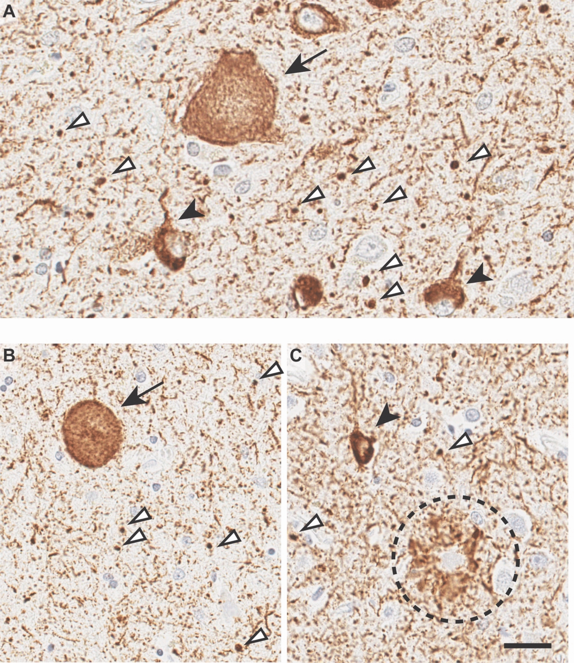
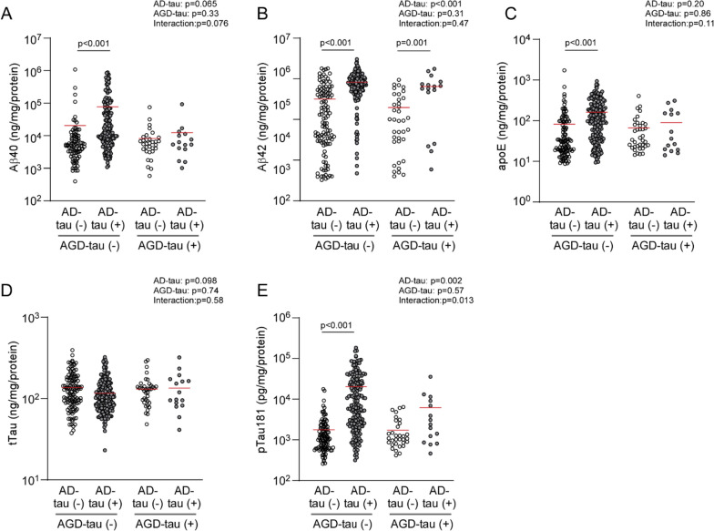
Alzheimer's disease (AD), characterized by the deposition of amyloid-β (Aβ) in senile plaques and neurofibrillary tangles of phosphorylated tau (pTau), is increasingly recognized as a complex disease with multiple pathologies. AD sometimes pathologically overlaps with age-related tauopathies such as four repeat (4R)-tau predominant argyrophilic grain disease (AGD). While AGD is often detected with AD pathology, the contribution of APOE4 to AGD risk is not clear despite its robust effects on AD pathogenesis. Specifically, how APOE genotype influences Aβ and tau pathology in co-occurring AGD and AD has not been fully understood. Using postmortem brain samples (N = 353) from a neuropathologically defined cohort comprising of cases with AD and/or AGD pathology built to best represent different APOE genotypes, we measured the amounts of major AD-related molecules, including Aβ40, Aβ42, apolipoprotein E (apoE), total tau (tTau), and pTau181, in the temporal cortex. The presence of tau lesions characteristic of AD (AD-tau) was correlated with cognitive decline based on Mini-Mental State Examination (MMSE) scores, while the presence of AGD tau lesions (AGD-tau) was not. Interestingly, while APOE4 increased the risk of AD-tau pathology, it did not increase the risk of AGD-tau pathology. Although APOE4 was significantly associated with higher levels of insoluble Aβ40, Aβ42, apoE, and pTau181, the APOE4 effect was no longer detected in the presence of AGD-tau. We also found that co-occurrence of AGD with AD was associated with lower insoluble Aβ42 and pTau181 levels. Overall, our findings suggest that different patterns of Aβ, tau, and apoE accumulation mediate the development of AD-tau and AGD-tau pathology, which is affected by APOE genotype.
Keywords: Alzheimer’s disease; Amyloid-β; Apolipoprotein E; Argyrophilic grain disease; MMSE; Tau.
© 2024. The Author(s).
Figures

References
-
- Das P, Verbeeck C, Minter L, Chakrabarty P, Felsenstein K, Kukar T, Maharvi G, Fauq A, Osborne BA, Golde TE. Transient pharmacologic lowering of Abeta production prior to deposition results in sustained reduction of amyloid plaque pathology. Mol Neurodegener. 2012;7:39. doi: 10.1186/1750-1326-7-39. - DOI - PMC - PubMed
Publication types
MeSH terms
Substances
Grants and funding
LinkOut - more resources
Full Text Sources
Medical
Miscellaneous
