. 2024 Feb 9;14(1):3333.
doi: 10.1038/s41598-024-53598-9.

Allogeneic umbilical cord blood-derived mesenchymal stem cell implantation versus microdrilling combined with high tibial osteotomy for cartilage regeneration

Affiliations

Allogeneic umbilical cord blood-derived mesenchymal stem cell implantation versus microdrilling combined with high tibial osteotomy for cartilage regeneration

Se-Han Jung et al. Sci Rep. .

Erratum in

Abstract

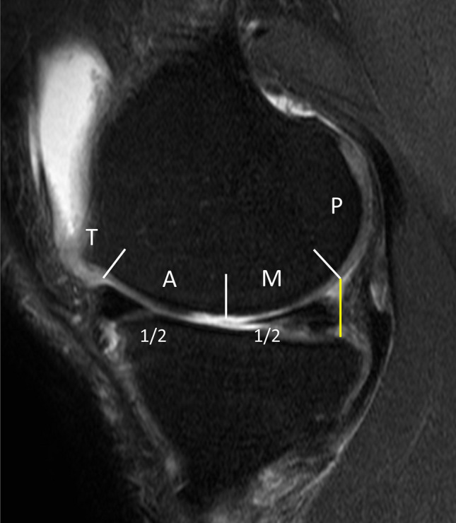
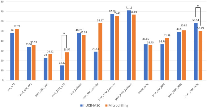
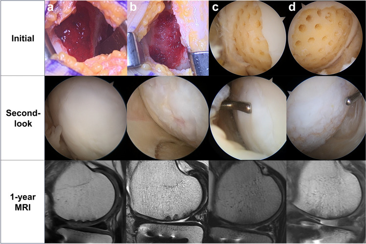
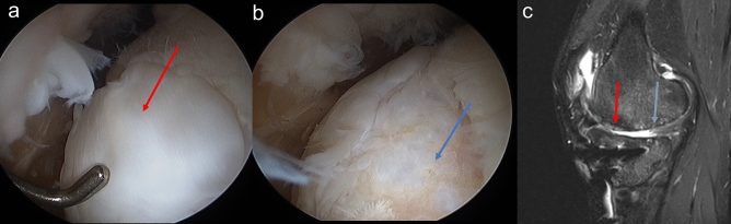
This study compared cartilage regeneration outcomes in knee osteoarthritis (OA) using allogeneic human umbilical cord blood-derived mesenchymal stem cell (hUCB-MSC) implantation and microdrilling with high tibial osteotomy (HTO). Fifty-four patients (60 knees) were included: 24 (27 knees) in the hUCB-MSC group and 30 (33 knees) in the microdrilling group. Both groups showed significant improvements in pain and functional scores at 6, 12, and 24 months compared to baseline. At 24 months, the hUCB-MSC group had significantly improved scores. Arthroscopic assessment at 12 months revealed better cartilage healing in the hUCB-MSC group. In subgroup analysis according to the defect site, hUCB-MSC implantation showed superior cartilage healing for anterior lesions. In conclusion, both treatments demonstrated effectiveness for medial OA. However, hUCB-MSC implantation had better patient-reported outcomes and cartilage regeneration than microdrilling. The study suggests promising approaches for cartilage restoration in large knee defects due to OA.


Conflict of interest statement

The authors declare no competing interests.

Figures

Figure 1
Figure 1
hUCB-MSC implantation and arthroscopic microdrilling procedures. (a) Arthroscopic view of the cartilage defect on medial femoral condyle viewed from the standard anterolateral portal in microdrilling group. (b) After arthroscopic microdrilling. (c) Exposure of the large cartilage defect on medial femoral condyle through mini-open arthrotomy. (d) A large cartilage defect extending anterior to posterior. (e) Drilling of the cartilage defect. (f) Implantation of the hUCB-MSCs.
Figure 2
Figure 2
Anatomical subdivision of the medial femoral condyle into trochlea (T), anterior (A), middle (M), and posterior (P) regions on sagittal projection.
Figure 3
Figure 3
Flowchart of patient inclusion in the study.
Figure 4
Figure 4
Patient-reported outcome scores across time points between groups. *Significant differences between groups with respect to scoring distributions at a specific time point in the student t-test at α = .05.
Figure 5
Figure 5
Cartilage regeneration outcomes in patients underwent two different procedures, hUCB-MSC implantation (a, b) and arthroscopic microdrilling (c, d). Initial, second-look arthroscopy, and postoperative MRI images depict the best (a, c) and the worst (b, d) cases among patients with anterior-to-mid lesions. (a) Fully covered medial femoral condyle (MFC) defect with cartilaginous tissue, achieving a MOCART score of 70. (b) Incomplete coverage of the MFC defect with irregular cobblestone appearance, resulting in a MOCART score of 35. (c) Complete coverage of the MFC defect after arthroscopic microdrilling, with softness on probing noted. (d) Minimal coverage of the MFC defect, presenting as soft and thin tissue, with a MOCART score of 25. MOCART, Magnetic Resonance Observation of Cartilage Repair Tissue.
Figure 6
Figure 6
Second-look arthroscopic findings of (a) the anterior lesion of MFC (ICRS Grade I) and (b) the mid to posterior lesion of MFC. (ICRS Grade IIIb) (c) At the 1-year follow-up, the anterior repaired cartilage (red arrow) completely filled the defect, but mid to posterior cartilage (blue arrow) showed an irregular surface and poor integration with incompletely filled defect at T2 PD FS MRI images.

References

    1. Biant LC, McNicholas MJ, Sprowson AP, Spalding T. The surgical management of symptomatic articular cartilage defects of the knee: consensus statements from United Kingdom knee surgeons. Knee. 2015;22:446–449. doi: 10.1016/j.knee.2015.06.001. - DOI - PubMed
    1. Seo SS, Kim CW, Jung DW. Management of focal chondral lesion in the knee joint. Knee. Surg. Relat. Res. 2011;23:185–196. doi: 10.5792/ksrr.2011.23.4.185. - DOI - PMC - PubMed
    1. Onoi Y, et al. Second-look arthroscopic findings and clinical outcomes after adipose-derived regenerative cell injection in knee osteoarthritis. Clin. Orthop. Surg. 2022;14:377–385. doi: 10.4055/cios20312. - DOI - PMC - PubMed
    1. Mina C, Garrett WE, Jr, Pietrobon R, Glisson R, Higgins L. High tibial osteotomy for unloading osteochondral defects in the medial compartment of the knee. Am. J. Sports Med. 2008;36:949–955. doi: 10.1177/0363546508315471. - DOI - PubMed
    1. Choi HU, Kim DH, Lee SW, Choi BC, Bae KC. Comparison of lower-limb alignment in patients with advanced knee osteoarthritis: EOS biplanar stereoradiography versus conventional scanography. Clin. Orthop. Surg. 2022;14:370–376. doi: 10.4055/cios21050. - DOI - PMC - PubMed