. 2024 Apr;8(4):651-662.
doi: 10.1038/s41559-024-02331-w. Epub 2024 Feb 9.

The Cabrières Biota (France) provides insights into Ordovician polar ecosystems

Affiliations

The Cabrières Biota (France) provides insights into Ordovician polar ecosystems

Farid Saleh et al. Nat Ecol Evol. 2024 Apr.

Abstract

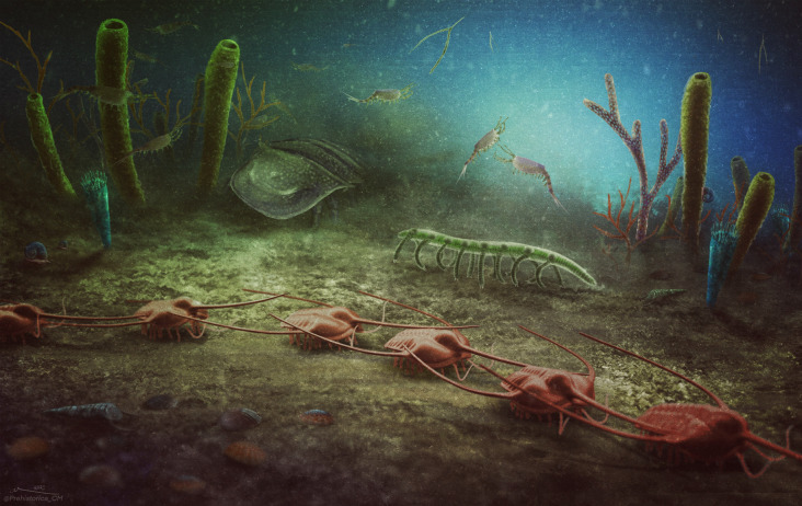
Early Palaeozoic sites with soft-tissue preservation are predominantly found in Cambrian rocks and tend to capture past tropical and temperate ecosystems. In this study, we describe the diversity and preservation of the Cabrières Biota, a newly discovered Early Ordovician Lagerstätte from Montagne Noire, southern France. The Cabrières Biota showcases a diverse polar assemblage of both biomineralized and soft-bodied organisms predominantly preserved in iron oxides. Echinoderms are extremely scarce, while sponges and algae are abundantly represented. Non-biomineralized arthropod fragments are also preserved, along with faunal elements reminiscent of Cambrian Burgess Shale-type ecosystems, such as armoured lobopodians. The taxonomic diversity observed in the Cabrières Biota mixes Early Ordovician Lagerstätten taxa with Cambrian forms. By potentially being the closest Lagerstätte to the South Pole, the Cabrières Biota probably served as a biotic refuge amid the high-water temperatures of the Early Ordovician, and shows comparable ecological structuring to modern polar communities.


Conflict of interest statement

The authors declare no competing interests.

Figures

Fig. 1
Fig. 1. Biomineralized taxa of the Cabrières Biota.
a, Trilobite of the genus Ampyx (UCBL-FSL713598). b, Gastropods associated with a tube-like structure, probably the conulariid Sphenothallus (UCBL-FSL713599). c, Biomineralized conulariid cnidarian (UCBL-FSL713600). d, Articulated brachiopods attached to a possible leptomitid sponge (UCBL-FSL713601). e, Assemblage formed of articulated brachiopods (centre), flattened carapaces probably of bivalved arthropods (centre left and right) and a calymenine trilobite cranidium (left; UCBL-FSL713602). f, A hyolith with possible internal organs (UCBL-FSL713603). Scale bars represent 4 mm in a and e, 1 cm in b and d, 5 mm in c, and 2 mm in f.
Fig. 2
Fig. 2. Sponges, algae and possible hemichordates from the Cabrières Biota.
a, A large sponge from the Cabrières Biota, possibly a demosponge (UCBL-FSL713604). b, Sponge (UCBL-FSL713605). c, Same specimen showing clear differentiation between the soft tissues (pink) and the mineralized skeleton (green) under multispectral imaging. d, Thick branching algae (UCBL-FSL713606). e, Thin branching algae (UCBL-FSL713607). f, More complex algae (UCBL-FSL713608). g, Organic tube of an Oesia-like enteropneust hemichordate (=Margaretia; UCBL-FSL713609). Scale bars represent 1 cm in a and f, 5 mm in e, and 3 mm in bd and g.
Fig. 3
Fig. 3. Non-biomineralized arthropods of the Cabrières Biota.
a, Phyllocarid carapace valves ornamented with very closely spaced, longitudinal striations and associated with abdominal segments (UCBL-FSL713609). b, Phyllocarid carapace valve with longitudinal striations preserved near a graptolite (UCBL-FSL713610). c, Possible chelicerate gnathobase (UCBL-FSL713611). d, Spiny arthropod appendage (UCBL-FSL713612). e, Segmented arthropod with chelicerate-like ornamentation and an eye (UCBL-FSL713613). f, Part of a segmented arthropod with chelicerate-like ornamentation and an appendage (UCBL-FSL713614). ap, appendage; co, chelicerate ornamentation; ey, eye; ls, longitudinal striations; sb, segmented body. Scale bars represent 2 mm in a; 8 mm in b, c and f; 5 mm in d and e; and 4 mm in j.
Fig. 4
Fig. 4. Vermiform organisms from the Cabrières Biota.
a, Unidentified vermiform organism UCBL-FSL713615, with possible remains of the gut and external ornamentation of tiny nodes. b,c, Incomplete armoured lobopodians UCBL-FSL713616 (b) and UCBL-FSL713617 (c) exhibiting two sclerite plates along an elongated soft body with annulations. A lateral extension in b possibly represents remains of the proximal part of a lobopod (?lo). d,e Close-up three-dimensional lateral (d) and top (e) views of part of UCBL-FSL713617, from the dotted box in c. Arrowheads point to lateral outgrowths associated with annulations that could represent spines or lobopod appendicules. an, annulations; gu, gut; lo, lobopod; otn, ornamentation of tiny nodes; ro, reticulate ornamentation; sp, sclerite plates. Scale bars represent 5 mm in a and c, and 1 mm in b and e; note that due to the three-dimensional rotation, no scale bar is given for d and the reader is instead invited to refer to scale bars in c and e.
Fig. 5
Fig. 5. Mode of fossil preservation.
a, Backscattered electron microscopy image revealing white iron oxide minerals and some limited black carbonaceous material within the fossils of the Cabrières Biota. b, The iron oxides appear shapeless, lacking distinct framboids or euhedral minerals. c, Semi-quantitative elemental proportions from SEM–EDX analyses indicate that the fossils exhibit a higher iron content compared with their surrounding aluminosilicate matrices. d, Fe K-edge XANES spectroscopy shows that the iron present in the Montagne Noire fossils exists in the form of oxides and hydroxides. e,f, Synchrotron µXRF major-to-trace elemental mapping shows that modern weathering elements such as manganese and arsenic are deposited on the surface of the samples. Scale bars represent 100 µm in a, 50 µm in b and 5 mm in e and f.
Fig. 6
Fig. 6. Artistic reconstruction of the Cabrières Biota.
In the foreground, a row of Ampyx (trilobites) and various shelly organisms, including brachiopods and a hyolith (bottom left corner). Behind the trilobites, a lobopodian, a chelicerate, cnidarians (blue), sponges (green), thin branching algae (red and green) and hemichordate tubes (purple), along with some molluscs. Bivalved arthropods inhabit the water column along with graptolites. Credit: Christian McCall (Prehistorica Art).
Extended Data Fig. 1
Extended Data Fig. 1. Distribution of Ordovician Lagerstätten.
(a) Early Ordovician palaeomap showing some of the significant Ordovician Lagerstätten, in addition to the newly discovered Cabrières Biota. (b) Stratigraphic formations yielding soft-tissue preservation during the Ordovician. The Cabrières Biota from the Landeyran Formation is the close to the Ordovician South Pole. It should be noted that some of the plotted sites in (a) date from the Middle or Upper Ordovician and not from the Early Ordovician. The paleomap is modified from Scotese but different reconstructions show rather comparable paleogeographic distributions. Dapingien (Dapin), Hirnantian (Hirn).
Extended Data Fig. 2
Extended Data Fig. 2. Geographic and stratigraphic context.
(a) Geographic position of the Cabrières Biota in the Montagne Noire in southern France, (b) within a 1 km radius around the Cabrières village. Map data ©2022 Google. The Cabrières Biota is discovered in the late Floian (Fl3) Landeyran Formation (c). (d) Photograph of the outcrop that yielded most soft-tissue preservation. (e) Geological map of the Montagne Noire showing the position of the new Lagerstätte in the southern part of the complex.
Extended Data Fig. 3
Extended Data Fig. 3. Taxonomic abundances in the Cabrières Biota and their comparison with the Early Ordovician Fezouata Biota.
The Cabrières Biota showcases a notably higher abundance of algae and sponges, while echinoderms are remarkably scarcer in comparison to the Fezouata Biota. Other animal taxa display relatively comparable representation between the two Lagerstätten. It is important to note that this data is highly probable to evolve with forthcoming fossil discoveries. Hence, it should be used only as an initial comparative reference. The Fezouata Biota dataset derives from the Marrakesh collections.
Extended Data Fig. 4
Extended Data Fig. 4. Additional views of the possible leptomitid sponge UCBL-FSL713601 shown in Fig. 1d.
(a) Optical photograph under polarised light of the entire specimen. (b) Close-up view under polarised light from the box in (a) showing projected longitudinal spicules. Scale bars represent 1 cm in (a), and 5 mm in (b).
Extended Data Fig. 5
Extended Data Fig. 5. Another possible leptomitid sponge, associated to the thin branching algae UCBL-FSL713607.
(a) Optical photograph under polarised light of the entire specimen. (b) Close-up view under polarised light from the box in (a) showing longitudinal skeletal elements. Scale bars represent 1 cm in (a), and 1 mm in (b).
Extended Data Fig. 6
Extended Data Fig. 6. Additional views of the large sponge UCBL-FSL713604 shown in Fig. 2a.
(a) Optical photograph of the entire organism. (b) Close-up view of one side of the organism, from the box in (a). (c) Close-up view of the osculum from the box in (b). (d, e) Close-up views under polarised light showing the skeletal framework, from the corresponding boxes in (c). Scale bars represent 1 cm in (a) and (b), 5 mm in (c), and 1 mm in (d) and (e).
Extended Data Fig. 7
Extended Data Fig. 7. Additional views of the algae shown in Fig. 2.
(a) Optical photograph of the thick branching algae UCBL-FSL713606 shown in Fig. 2d. (b) Close-up view under polarised light of basal branches, from the box in (a). (c) Optical photograph under polarised light of the thin branching algae UCBL-FSL713607 shown in Fig. 2e. (d) Close-up view under polarised light from the box in (c) highlighting textural differences between algae (right) and sponges (left). (e) Optical photograph under low angle and polarised light of the more complex algae UCBL-FSL713608 shown in Fig. 2f. (f) Close-up view under polarised light from the box in (e) highlighting branched clumps of flat kidney-shaped segments. Scale bars represent 5 cm in (a), (b), (c) and (f), 1 mm in (d), and 1 cm in (e).
Extended Data Fig. 8
Extended Data Fig. 8. Phyllocarid crustaceans from the Cabrières Biota.
(a, b) Optical photograph (a) and interpretative line drawing (b) of the counterpart of UCBL-FSL713609 shown in Fig. 3a. The specimen contains two left valves ornamented with very closely spaced, longitudinal striations. The bottom valve (white) is associated with a few abdominal segments. Fossil remains located anteriorly to the top valve (light blue) may represent a rostral plate. (c, d) Optical photograph (c) and interpretative line drawing (d) of the left valve UCBL-FSL713610 shown in Fig. 3b. Abbreviations: al, anterodorsal lobe; as, abdominal segments; cc, cephalic carina; co, carapace ornamentation; mdp, median dorsal plate; rp, rostral plate. Scale bars represent 5 mm.
Extended Data Fig. 9
Extended Data Fig. 9. Additional images and interpretative drawings of the chelicerates in Fig. 3e, f.
(a, b) Optical photograph (a) and interpretative line drawing (b) of the chelicerate UCBL-FSL713613. The presence of a prosoma lacking enlarged axial nodes on opisthosomal tergites, reduced genal spines, and an opisthosoma that is largest at the third and fourth tergite, likely suggest chasmataspid affinities. (c) Close up of the eye and the ornamentation. (dg) Optical photographs under polarised light of the part (d) and the counterpart (f), and corresponding interpretative line drawings (e, g) of the chelicerate UCBL-FSL713614. Green highlights ventral features. Pale purple shows dorsal features. The sample displays a carapace, three tergites, and a possible appendage. The presence of a possible metastoma suggest non-arachnid dekatriatan affinities. (h, i) Close-up photographs showing the ornamentation. Abbreviations: appendage (ap); (ax) axis; cardiac lobe (cdl); doublure (db); ornamentation (or); eye (ey); metastoma (m); and tergite (t). Scale bars represent 2 mm in (c)s and (i), 3 mm in (h), 5 mm in (a) and (b), and 8 mm in (d), (e), (f), and (g).
Extended Data Fig. 10
Extended Data Fig. 10. Synchrotron-based X-ray fluorescence major-to-elemental mapping of the Cabrières Biota.
(a) Optical photograph under polarised light of the assemblage UCBL-FSL713602 shown in Fig. 1e. (b, c) False-colour overlays of manganese (red), iron (green) and potassium (blue) (b), and arsenic (red), iron (green) and potassium (blue) (c) distributions from the box in (a). Acquisition parameters: 200 μm steps, 80 ms dwell time, 51,100 pixels. (d) Optical photograph under polarised light of sponge UCBL-FSL713618. (e) Close-up view from the box in (d). (f, g) False-colour overlays of manganese (red), iron (green) and potassium (blue) distributions from the corresponding boxes in (e) and (f). Acquisition parameters: 200 μm steps, 60 ms dwell time, 6,150 pixels in (f); 100 μm steps, 60 ms dwell time, 4,800 pixels in (g). The white circle in (g) locates the XANES analysis shown in Fig. 4d. (h) Average µXRF spectra and main elemental contributions from the corresponding numbered boxes in c (156 pixels) and f (208 pixels). Scale bars represent 1 cm in (a), (b), (c) and (d), 5 mm in (e) and (f), and 1 mm in (g).

References

    1. Seilacher, A., Reif, W. E. & Westphal, F. Sedimentological, ecological and temporal patterns of fossil Lagerstätten. Philos. Trans. R. Soc. Lond. B311, 5–24 (1985).
    1. Allison, P. A. & Briggs, D. E. Exceptional fossil record: distribution of soft-tissue preservation through the Phanerozoic. Geology21, 527–530 (1993).
    1. Brasier, M. D., Antcliffe, J. B. & Callow, R. H. T. in Taphonomy: Process and Bias Through Time 2nd edn, Vol. 32 (eds P. A. Allison & D. J. Bottjer) Ch. 15, 519–567 (Springer Science, 2011).
    1. Muscente, A. D. et al. Exceptionally preserved fossil assemblages through geologic time and space. Gondwana Res.48, 164–188 (2017).
    1. Van Roy, P. et al. Ordovician faunas of Burgess Shale type. Nature465, 215–218 (2010). - PubMed