. 2024 Jan 30;16(3):379.
doi: 10.3390/polym16030379.

Features of Changes in the Structure and Properties of a Porous Polymer Material with Antibacterial Activity during Biodegradation in an In Vitro Model

Affiliations

Features of Changes in the Structure and Properties of a Porous Polymer Material with Antibacterial Activity during Biodegradation in an In Vitro Model

Vladimir V Yudin et al. Polymers (Basel). .

Abstract

Hybrid porous polymers based on poly-EGDMA and polylactide containing vancomycin, the concentration of which in the polymer varied by two orders of magnitude, were synthesized. The processes of polymer biodegradation and vancomycin release were studied in the following model media: phosphate-buffered saline (PBS), trypsin-Versene solution, and trypsin-PBS solution. The maximum antibiotic release was recorded during the first 3 h of extraction. The duration of antibiotic escape from the polymer samples in trypsin-containing media varied from 3 to 22 days, depending on the antibiotic content of the polymer. Keeping samples of the hybrid polymer in trypsin-containing model media resulted in acidification of the solutions-after 45 days, up to a pH of 1.84 in the trypsin-Versene solution and up to pH 1.65 in the trypsin-PBS solution. Here, the time dependences of the vancomycin release from the polymer into the medium and the decrease in pH of the medium correlated. These data are also consistent with the results of a study of the dynamics of sample weight loss during extraction in the examined model media. However, while the polymer porosity increased from ~53 to ~60% the pore size changed insignificantly, over only 10 μm. The polymer samples were characterized by their antibacterial activity against Staphylococcus aureus, and this activity persisted for up to 21 days during biodegradation of the material, regardless of the medium type used in model. Surface-dependent human cells (dermal fibroblasts) adhere well, spread out, and maintain high viability on samples of the functionalized hybrid polymer, thus demonstrating its biocompatibility in vitro.

Keywords: antibiotic release; cell adhesion; ethylene glycol dimethacrylate; photopolymerization; polylactide; porosity; porous polymer; scaffolds; scanning electron microscopy.


Conflict of interest statement

The authors declare no conflicts of interest.

Figures

Figure 1
Figure 1
Change in vancomycin content in the polymer sample extracts (accumulated over 3 h extraction periods) with different drug contents, depending on the overall exposure time in phosphate-buffered saline.
Figure 2
Figure 2
Vancomycin content in extracts of polymer samples with different concentrations of the antibiotic when kept in a trypsin-Versene solution and sampled at the specified intervals.
Figure 3
Figure 3
Vancomycin content in extracts of polymer samples with different drug contents when kept in the trypsin-PBS solution and sampled at the specified intervals.
Figure 4
Figure 4
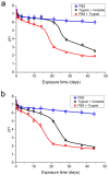
Change in the pH of the model media of the samples depending on the time of exposure: (a) samples without vancomycin; (b) samples with 10% vancomycin.
Figure 5
Figure 5
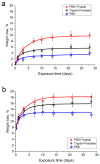
Dependence of the relative weight loss of the hybrid polymer samples (a) without vancomycin and (b) with vancomycin (10%) on the duration of the samples’ exposure to the model solutions: (a) weight losses from samples relative to the weight of polylactide in the samples; (b) weight losses from samples relative to the total weight of polylactide and vancomycin in the samples.
Figure 6
Figure 6
SEM images of fractures: samples before exposure to a physiological medium—porous polymer matrix (a) before (sample No. 1) and (b) after treatment with the PLA solution (sample No. 2), as well as (c) after treatment with the PLA solution with vancomycin (sample No. 3); samples of porous polymer materials treated with the PLA solution and kept in a phosphate-buffered saline for 28 days (d) (sample No. 6); in the trypsin-Versene solution (e) (sample No. 9) and a mixture of phosphate buffer and trypsin (f) (sample No. 12); samples of porous polymeric materials treated with the PLA solution with vancomycin and kept in phosphate-buffered saline for 28 days (g) (sample No. 15), in the trypsin-Versene solution (h) (sample No. 19) and the trypsin-PBS solution (i) (sample No. 23).
Figure 6
Figure 6
SEM images of fractures: samples before exposure to a physiological medium—porous polymer matrix (a) before (sample No. 1) and (b) after treatment with the PLA solution (sample No. 2), as well as (c) after treatment with the PLA solution with vancomycin (sample No. 3); samples of porous polymer materials treated with the PLA solution and kept in a phosphate-buffered saline for 28 days (d) (sample No. 6); in the trypsin-Versene solution (e) (sample No. 9) and a mixture of phosphate buffer and trypsin (f) (sample No. 12); samples of porous polymeric materials treated with the PLA solution with vancomycin and kept in phosphate-buffered saline for 28 days (g) (sample No. 15), in the trypsin-Versene solution (h) (sample No. 19) and the trypsin-PBS solution (i) (sample No. 23).
Figure 7
Figure 7
The sizes of the growth inhibition areas on staphylococcus test cultures around samples of the control disk with vancomycin (No. 1) and samples of the hybrid polymer with a vancomycin concentration of 10% after immersion in phosphate-buffered saline (No. 2), in the trypsin-PBS solution (No. 3) and in the trypsin-Versene solution (No. 4), depending on the time in the medium: 3 h (a), 1 day (b), 3 days (c), 7 days (d), 14 days (e) and 21 days (f).
Figure 7
Figure 7
The sizes of the growth inhibition areas on staphylococcus test cultures around samples of the control disk with vancomycin (No. 1) and samples of the hybrid polymer with a vancomycin concentration of 10% after immersion in phosphate-buffered saline (No. 2), in the trypsin-PBS solution (No. 3) and in the trypsin-Versene solution (No. 4), depending on the time in the medium: 3 h (a), 1 day (b), 3 days (c), 7 days (d), 14 days (e) and 21 days (f).
Figure 8
Figure 8
Fibroblasts on the surface of the hybrid polymer samples: (a) fibroblast nuclei on the surface of the hybrid polymer samples, stained blue (Hoechst fluorochrome); (b) fibroblasts spread on the surface of the hybrid polymer samples: the cell cytoplasm is stained green (Calcein AM fluorochrome), and cell nuclei are blue (Hoechst fluorochrome).

Similar articles

Cited by

References

    1. Kovylin R., Aleynik D.Y., Fedushkin I. Modern Porous Polymer Implants: Synthesis, Properties, and Application. Polym. Sci. Ser. C. 2021;63:29–46. doi: 10.1134/S1811238221010033. - DOI
    1. Chesnokov S.A., Aleynik D.Y., Kovylin R.S., Yudin V.V., Egiazaryan T.A., Egorikhina M.N., Zaslavskaya M.I., Rubtsova Y.P., Gusev S.A., Mlyavykh S.G. Porous Polymer Scaffolds based on Cross-Linked Poly-EGDMA and PLA: Manufacture, Antibiotics Encapsulation, and In Vitro Study. Macromol. Biosci. 2021;21:2000402. doi: 10.1002/mabi.202000402. - DOI - PubMed
    1. Kwon D.Y., Park J.Y., Lee B.Y., Kim M.S. Comparison of scaffolds fabricated via 3D printing and salt leaching: In vivo imaging, biodegradation, and inflammation. Polymers. 2020;12:2210. doi: 10.3390/polym12102210. - DOI - PMC - PubMed
    1. Velasco M.A., Narváez-Tovar C.A., Garzón-Alvarado D.A. Design, materials, and mechanobiology of biodegradable scaffolds for bone tissue engineering. BioMed Res. Int. 2015;2015:1–21. doi: 10.1155/2015/729076. - DOI - PMC - PubMed
    1. Kim J.-A., Lim J., Naren R., Yun H.-s., Park E.K. Effect of the biodegradation rate controlled by pore structures in magnesium phosphate ceramic scaffolds on bone tissue regeneration in vivo. Acta Biomater. 2016;44:155–167. doi: 10.1016/j.actbio.2016.08.039. - DOI - PubMed

LinkOut - more resources