Review
. 2024 Jan 26;13(3):717.
doi: 10.3390/jcm13030717.

Right Ventricular Function in Arrhythmogenic Right Ventricular Cardiomyopathy: Potential Value of Strain Echocardiography

Affiliations
Review

Right Ventricular Function in Arrhythmogenic Right Ventricular Cardiomyopathy: Potential Value of Strain Echocardiography

Caroline Løkke Bjerregaard et al. J Clin Med. .

Abstract

Arrhythmogenic right ventricular cardiomyopathy is an inherited cardiomyopathy, characterized by abnormal cell adhesions, disrupted intercellular signaling, and fibrofatty replacement of the myocardium. These changes serve as a substrate for ventricular arrhythmias, placing patients at risk of sudden cardiac death, even in the early stages of the disease. Current echocardiographic criteria for diagnosing arrhythmogenic right ventricular cardiomyopathy lack sensitivity, but novel markers of cardiac deformation are not subject to the same technical limitations as current guideline-recommended measures. Measuring cardiac deformation using speckle tracking allows for meticulous quantification of global systolic function, regional function, and dyssynchronous contraction. Consequently, speckle tracking to quantify myocardial strain could potentially be useful in the diagnostic process for the determination of disease progression and to assist risk stratification for ventricular arrhythmias and sudden cardiac death. This narrative review provides an overview of the potential use of different myocardial right ventricular strain measures for characterizing right ventricular dysfunction in arrhythmogenic right ventricular cardiomyopathy and its utility in assessing the risk of ventricular arrhythmias.

Keywords: ARVC; arrhythmogenic cardiomyopathy; right ventricle; speckle tracking; strain.


Conflict of interest statement

C.L.B.: none; T.B.-S.: research grants from Sanofi Pasteur, GSK, Novo Nordisk, AstraZeneca, Boston Scientific, and GE Healthcare; consulting fees from Novo Nordisk, IQVIA, Parexel, Amgen, CSL Seqirus, GSK, and Sanofi Pasteur; and lecture fees from Bayer, Novartis, Sanofi Pasteur, GE Healthcare, and GSK. K.G.S.: advisory board member at Sanofi Pasteur. M.S.: none. M.C.H.L.: none. N.D.J.: none. F.J.O.: none. The organizations had no role in any part of this review.

Figures
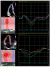
Figure 1
Figure 1
Representative examples of RV speckle tracking. Both figures represent speckle tracking performed in the modified 4-chamber, RV-focused view. (A) is an example of a healthy individual. Segmental strain curves are shown as colored lines, whereas the global value is shown as white dotted line. Note the global strain value of −21.6% and the synchronous contraction pattern with peak strain values aligned at the green line (corresponding to pulmonic valve closure). (B) is an example of speckle tracking in a patient with overt ARVC. Note the lower global strain value of −17.4%, the heterogenous contraction pattern, and delayed onset of contraction with post-systolic shortening in the subctricuspid segment (yellow curve). RV: right ventricular; ARVC: arrhythmogenic right ventricular cardiomyopathy.
Figure 2
Figure 2
RV deformation pattern classification. Based on the presence of different strain abnormalities in the right ventricular (RV) basal area, 3 distinct characteristic RV deformation patterns were identified: type I represents normal deformation; type II shows delayed onset of shortening (yellow dot) and reduced systolic peak strain (white square) compared with normal deformation and presence of post-systolic shortening (arrow); and type III displays prominent systolic stretching (upward arrows) and passive recoil or shortening (downward arrow) during early diastole. Dotted white lines indicate type I deformation pattern for comparison. PVO/PVC 1⁄4 pulmonary valve opening/closure” Mast et al. [52]. Reproduced with permission from RightsLink/Elsevier.

Similar articles

Cited by

References

    1. Marcus F.I., Fontaine G.H., Guiraudon G., Frank R., Laurenceau J.L., Malergue C., Grosgogeat Y. Right Ventricular Dysplasia: A Report of 24 Adult Cases. Circulation. 1982;65:384–398. doi: 10.1161/01.CIR.65.2.384. - DOI - PubMed
    1. Corrado D., Basso C., Judge D.P. Arrhythmogenic Cardiomyopathy. Circ. Res. 2017;121:784–802. doi: 10.1161/CIRCRESAHA.117.309345. - DOI - PubMed
    1. Corrado D., Zorzi A., Cipriani A., Bauce B., Bariani R., Beffagna G., De Lazzari M., Migliore F., Pilichou K., Rampazzo A., et al. Evolving Diagnostic Criteria for Arrhythmogenic Cardiomyopathy. J. Am. Heart Assoc. 2021;10:e021987. doi: 10.1161/JAHA.121.021987. - DOI - PMC - PubMed
    1. Corrado D., Link M.S., Calkins H. Arrhythmogenic Right Ventricular Cardiomyopathy. N. Engl. J. Med. 2017;376:61–72. doi: 10.1056/NEJMra1509267. - DOI - PubMed
    1. Arbelo E., Protonotarios A., Gimeno J.R., Arbustini E., Barriales-Villa R., Basso C., Bezzina C.R., Biagini E., Blom N.A., de Boer R.A., et al. 2023 ESC Guidelines for the Management of Cardiomyopathies. Eur. Heart J. 2023;44:3503–3626. doi: 10.1093/eurheartj/ehad194. - DOI - PubMed

LinkOut - more resources