

The Prognostic Role of Volumetric MRI Evaluation in the Surgical Treatment of Glioblastoma
- PMID: 38337543
- PMCID: PMC10856584
- DOI: 10.3390/jcm13030849
The Prognostic Role of Volumetric MRI Evaluation in the Surgical Treatment of Glioblastoma
Abstract
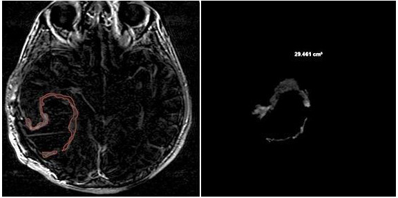
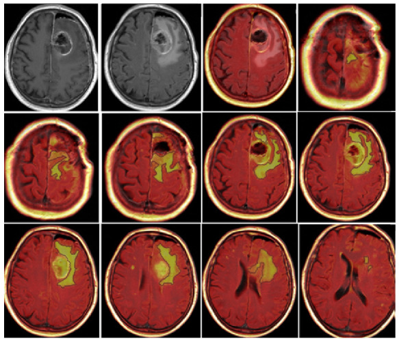
Background: Glioblastoma is the most common primary brain neoplasm in adults, with a poor prognosis despite a constant effort to improve patient survival. Some neuroradiological volumetric parameters seem to play a predictive role in overall survival (OS) and progression-free survival (PFS). The aim of this study was to analyze the impact of the volumetric areas of contrast-enhancing tumors and perineoplastic edema on the survival of patients treated for glioblastoma. Methods: A series of 87 patients who underwent surgery was retrospectively analyzed; OS and PFS were considered the end points of the study. For each patient, a multidisciplinary revision was conducted in collaboration with the Neuroradiology and Neuro-Oncology Board. Manual and semiautomatic measurements were adopted to perform the radiological evaluation, and the following quantitative parameters were retrospectively analyzed: contrast enhancement preoperative tumor volume (CE-PTV), contrast enhancement postoperative tumor volume (CE-RTV), edema/infiltration preoperative volume (T2/FLAIR-PV), edema/infiltration postoperative volume (T2/FLAIR-RV), necrosis volume inside the tumor (NV), and total tumor volume including necrosis (TV). Results: The median OS value was 9 months, and the median PFS value was 4 months; the mean values were 12.3 and 6.9 months, respectively. Multivariate analysis showed that the OS-related factors were adjuvant chemoradiotherapy (p < 0.0001), CE-PTV < 15 cm3 (p = 0.03), surgical resection > 95% (p = 0.004), and the presence of a "pseudocapsulated" radiological morphology (p = 0.04). Conclusions: Maximal safe resection is one of the most relevant predictive factors for patient survival. Semiautomatic preoperative MRI evaluation could play a key role in prognostically categorizing these tumors.
Keywords: FLAIR infiltration; brain tumors; extent of surgical resection; glioblastoma; neuro-oncology; overall survival; progression-free survival; pseudocapsule; tumor volume.
Conflict of interest statement
The authors declare no conflicts of interest.
Figures


Similar articles
-
Residual tumor volume versus extent of resection: predictors of survival after surgery for glioblastoma.J Neurosurg. 2014 Nov;121(5):1115-23. doi: 10.3171/2014.7.JNS132449. Epub 2014 Sep 5. J Neurosurg. 2014. PMID: 25192475
-
Apparent diffusion coefficient and tumor volume measurements help stratify progression-free survival of bevacizumab-treated patients with recurrent glioblastoma multiforme.Neuroradiol J. 2019 Aug;32(4):241-249. doi: 10.1177/1971400919847184. Epub 2019 May 8. Neuroradiol J. 2019. PMID: 31066622 Free PMC article.
-
Dynamics of FLAIR Volume Changes in Glioblastoma and Prediction of Survival.Ann Surg Oncol. 2017 Mar;24(3):794-800. doi: 10.1245/s10434-016-5635-z. Epub 2016 Oct 20. Ann Surg Oncol. 2017. PMID: 27766560
-
Volumetric quantification of glioblastoma: experiences with different measurement techniques and impact on survival.J Neurooncol. 2017 Nov;135(2):391-402. doi: 10.1007/s11060-017-2587-5. Epub 2017 Jul 28. J Neurooncol. 2017. PMID: 28755324 Review.
-
Cerebellar glioblastoma: a retrospective review of 21 patients at a single institution.J Neurooncol. 2011 Dec;105(3):555-62. doi: 10.1007/s11060-011-0617-2. Epub 2011 Jun 4. J Neurooncol. 2011. PMID: 21643841 Review.
Cited by
-
Diagnostic Utility of Intratumoral Susceptibility Signals in Adult Diffuse Gliomas: Tumor Grade Prediction and Correlation with Molecular Markers Within the WHO CNS5 (2021) Classification.J Clin Med. 2025 Jun 5;14(11):4004. doi: 10.3390/jcm14114004. J Clin Med. 2025. PMID: 40507765 Free PMC article.
-
Glioblastoma: From Pathophysiology to Novel Therapeutic Approaches.Biomedicines. 2025 Aug 12;13(8):1963. doi: 10.3390/biomedicines13081963. Biomedicines. 2025. PMID: 40868217 Free PMC article. Review.
-
Focused ultrasound as a treatment modality for gliomas.Front Neurol. 2024 May 15;15:1387986. doi: 10.3389/fneur.2024.1387986. eCollection 2024. Front Neurol. 2024. PMID: 38813245 Free PMC article. Review.
References
-
- Stoyanov G.S., Lyutfi E., Georgieva R., Georgiev R., Dzhenkov D.L., Petkova L., Ivanov B.D., Kaprelyan A., Ghenev P. Reclassification of Glioblastoma Multiforme According to the 2021 World Health Organization Classification of Central Nervous System Tumors: A Single Institution Report and Practical Significance. Cureus. 2022;14:e21822. doi: 10.7759/cureus.21822. - DOI - PMC - PubMed
LinkOut - more resources
Full Text Sources
