Review
. 2024 Feb 2;13(3):876.
doi: 10.3390/jcm13030876.

Approach to Acute Myeloid Leukemia with Increased Eosinophils and Basophils

Affiliations
Review

Approach to Acute Myeloid Leukemia with Increased Eosinophils and Basophils

Stavros Papadakis et al. J Clin Med. .

Abstract

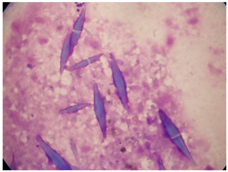
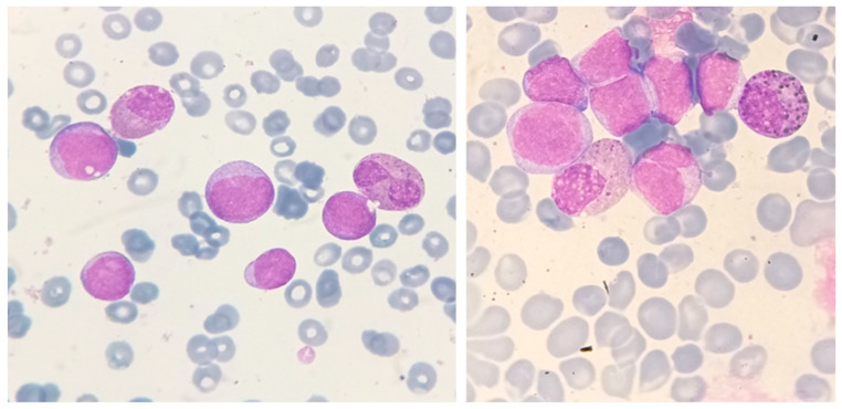
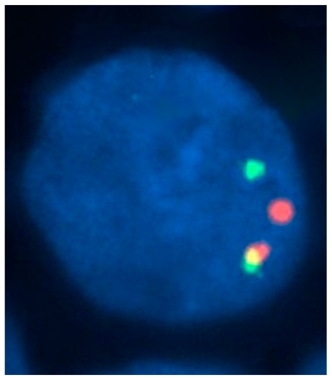
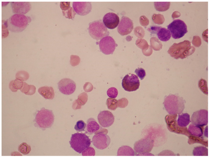
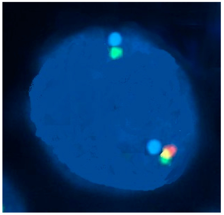
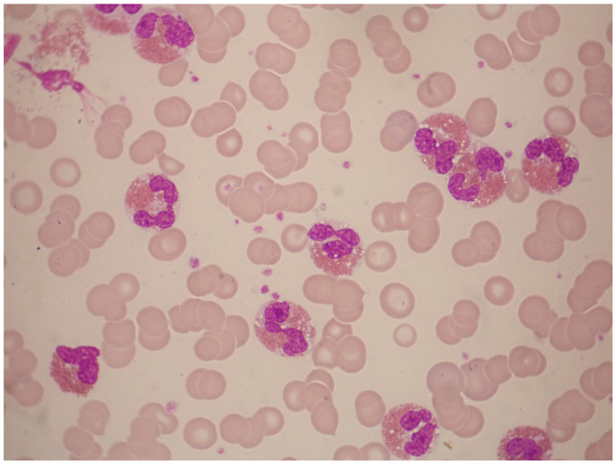
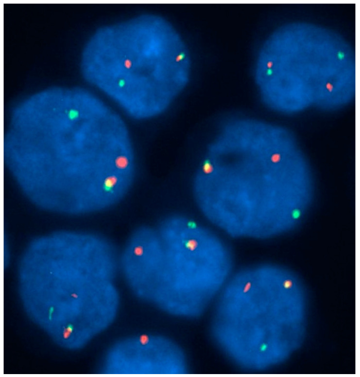
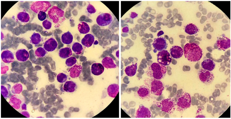
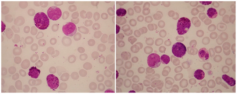
There is remarkable morphologic and genetic heterogeneity in acute myeloid leukemia (AML). In a small percentage of cases of AML, increased eosinophils and/or basophils are present in the bone marrow and sometimes in the peripheral blood. This is often a puzzling diagnostic situation but also an important finding that requires special investigation. Unique chromosomal rearrangements have been correlated with an increased number of eosinophils and basophils in AML. The identification of the underlying genetic lesion that promotes eosinophilia and basophilia can dramatically change both the prognosis and the treatment of the patient. Thus, clinicians must be vigilant in searching for the cause of eosinophilia and basophilia in patients with AML, since the different causes may lead to different treatments and survival outcomes. In this article, we examine the significance of increased eosinophils and/or basophils in the context of AML, provide guidance that simplifies the differential diagnosis, and give prognostic and therapeutic information about specific subtypes of AML associated with eosinophilia and/or basophilia. Evidence supporting personalized (molecularly targeted) therapy for these patients is also presented.

Keywords: AML; CBF; PDGFRA; PDGFRB; acute myeloid leukemia; basophil; basophilia; eosinophil; eosinophilia; morphology.


Conflict of interest statement

The authors declare no conflicts of interest.

Figures

Figure 1
Figure 1
Charcot–Leyden crystals in association with AML. These bizarre bipyramidal crystal structures were noted in the bone marrow aspirate smears of a 75-year-old woman with pancytopenia.
Figure 2
Figure 2
Abnormal eosinophils in the bone-marrow aspirates of a patient with AML with inv(16)(p13.1q22). These micrographs, showing abnormal eosinophils containing both eosinophilic and basophilic staining granules, are representative of myelomonocytic leukemia with eosinophilia (May–Grünwald–Giemsa, ×1000).
Figure 3
Figure 3
An interphase fluorescence in situ hybridization (FISH) study with a dual-color break-apart probe set containing two probes flanking the breakpoint in the CBFB gene, showing a nucleus with inv(16)(p13.1 q22). An intact gene results in the colocalization of the two probes, producing a fusion (yellow) signal. The presence of CBFB rearrangement is indicated by separate red and green signals.
Figure 4
Figure 4
Bone-marrow aspirate smear of a 54-year-old man with AML with t(8;21)(q22;q22) translocation and eosinophilia. The white-cell count was 2.0 × 10⁹/L, without increased eosinophils in the peripheral blood. In the bone marrow, however, eosinophils and eosinophil precursors constituted 22% of cells, without abnormal basophilic granules (May–Grünwald–Giemsa, ×1000).
Figure 5
Figure 5
Detection of FIP1L1-PDGFRA in a case of AML with eosinophilia, using a three-color probe strategy. On interphase fluorescence in situ hybridization performed with the use of probes to LNX1 (in red), FIP1L1 (in green), and PDGFRA (in aqua), the nucleus shows one chromosome with all three signals intact and another chromosome with intact FIP1L1 and PDGFRA signals but without the LNX1 signal (i.e., loss of red signal). This finding indicates a deletion of the region between FIP1L1 and PDGFRA on chromosome 4q12, consistent with the FIP1L1-PDGFRA fusion.
Figure 6
Figure 6
Abnormal eosinophil morphology associated with FIP1L1-PDGFRA rearrangement. This peripheral-blood smear shows eosinophils with trilobed nuclei or hypersegmented eosinophils as well as eosinophils with many cytoplasmic vacuoles due to degranulation (May–Grünwald–Giemsa stain, ×1000).
Figure 7
Figure 7
A 70-year-old man with monocytosis and eosinophilia due to PDGFRB rearrangement. An interphase fluorescence in situ hybridization study with a dual-color break-apart probe set containing two probes flanking the sequence of PDGFRB gene showed that most cells had one fused signal and separate red and green signals, indicating disruption of the PDGFRB gene (a normal gene produces a colocalization, i.e., a yellow signal, whereas a rearranged gene results in two separate green and red signals).
Figure 8
Figure 8
Acute mast-cell leukemia. Bone marrow aspirate smears of a 79-year-old man with aleukemic acute mast-cell leukemia who presented with pancytopenia and abnormal liver function tests. The photomicrographs show round neoplastic cells with dark cytoplasmic granules. The cells were CD13+, HLA-DR+, CD33+, CD203c+, CD38+, CD2, CD9, CD123, CD11b, FcεRI+, CD117+, and CD25+. PCR was positive for C-KIT D816V mutation.
Figure 9
Figure 9
Illustrative case of acute basophilic leukemia (ABL). A 67-year-old man presented for evaluation of general weakness and lower back pain. Over the previous 8 months, he had had postprandial epigastric pain. He had undergone esophagogastroduodenoscopy, which showed the presence of three simultaneous gastric ulcers. Biopsies were negative for Helicobacter pylori infection. His past medical history was otherwise unremarkable. He smoked 20 cigarettes daily, drank alcohol rarely, and took no medications. On examination, there was mild hepatomegaly (2 cm below right coastal margin), small-volume peripheral lymphadenopathy (diameter, ≤2 cm), and no splenomegaly. Multiple skin lesions were noted on the trunk and extremities, measuring a few centimeters. They were reddish, reddish brown, or purple, plaque-like, and produced significant itching and discomfort. A complete blood count revealed anemia (hemoglobin, 9.5 g/dL; ΜCV 80 fL), white-cell count 5.1 × 10⁹/L, and platelet count 45 × 109/L. May–Grünwald–Giemsa staining of a peripheral-blood smear revealed 41% neutrophils, 36% lymphocytes, 1% monocytes, 8% nucleated red blood cells, and 22% “atypical” cells with basophilic granules, easily identifiable under oil immersion (×1000). Coagulation studies and hepatic biochemistry were normal, but there was renal dysfunction (urea; 65 mg/dL, creatinine: 2.2 mg/dL) and hypocalcemia (6.2 mg/dL). Lactate dehydrogenase level was 1376 U/L (<246 U/L). Flow cytometry showed that the immature cell population in the CD45weak/SSClow gate was CD34+, MPO, CD11b+, CD13+, CD33+, CD9+, CD123+, CD203c+, HLA-DR, CD2, CD3, CD5, CD16, CD10, CD22weak, CD25weak, CD15, TdT, and CD117. An attempted aspiration of the bone marrow yielded a "dry tap”. The most striking feature in this patient was the presence of blast cells with coarse basophilic granules, raising suspicion for ABL. ABL can be identified by expression of either CD123 or CD203c by cells that do not express CD117. The absence of myeloperoxidase (MPO) rules out the possibility of acute promyelocytic leukemia (M3b). In this case, positivity for CD11b and CD123 and the absence of CD117 strongly suggested a diagnosis of ABL. PML-RARA, BCR-ABL1, RUNX1-RUNX1T1, CBFβ-MYH11, and C-KIT D816V were negative. NPM1 and FLT3-ITD, FLT3-TKD mutations were also negative. A peripheral-blood sample for cytogenetic analysis showed no abnormalities (46, XY). The patient’s symptoms can be explained on the basis of hyperhistaminemia. Accordingly, the histamine levels were found to be 710 pg/mL (normal, 0–90 pg/mL). The patient was treated with high doses of two H1 inhibitors, an H2 inhibitor, esomeprazole, and methylprednisone, before initiation of induction chemotherapy.

Similar articles

Cited by

References

    1. Metcalf D. Hematopoietic regulators: Redundancy or subtlety? Blood. 1993;82:3515–3523. doi: 10.1182/blood.V82.12.3515.3515. - DOI - PubMed
    1. Mack E.A., Pear W.S. Transcription factor and cytokine regulation of eosinophil lineage commitment. Curr. Opin. Hematol. 2020;27:27–33. doi: 10.1097/MOH.0000000000000552. - DOI - PMC - PubMed
    1. Tenen D.G. Disruption of differentiation in human cancer: AML shows the way. Nat. Rev. Cancer. 2003;3:89–101. doi: 10.1038/nrc989. - DOI - PubMed
    1. Kato M., Kephart G.M., Talley N.J., Wagner J.M., Sarr M.G., Bonno M., McGovern T.W., Gleich G.J. Eosinophil infiltration and degranulation in normal human tissue. Anat. Rec. 1998;252:418–425. doi: 10.1002/(SICI)1097-0185(199811)252:3<418::AID-AR10>3.0.CO;2-1. - DOI - PubMed
    1. Hoffman R., Benz E., Silberstein L., Heslop H., Weitz J., Anastasi J., Salama M. Hematology: Basic Principles and Practice. Elsevier; Philadelphia, PA, USA: 2018. pp. 330–331. - DOI

LinkOut - more resources