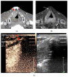
The Diagnostic Value of CEUS in Assessing Non-Ossified Thyroid Cartilage Invasion in Patients with Laryngeal Squamous Cell Carcinoma
- PMID: 38337585
- PMCID: PMC10856113
- DOI: 10.3390/jcm13030891
The Diagnostic Value of CEUS in Assessing Non-Ossified Thyroid Cartilage Invasion in Patients with Laryngeal Squamous Cell Carcinoma
Abstract
Background: Accurate assessment of thyroid cartilage invasion in squamous cell carcinoma (SCC) of the larynx remains a challenge in clinical practice. The aim of this study was to assess the diagnostic performance of contrast-enhanced ultrasound (CEUS), contrast-enhanced computed tomography (CECT), and magnetic resonance imaging (MRI) in the detection of non-ossified thyroid cartilage invasion in patients with SCC. Methods: CEUS, CECT, and MRI scans of 27 male patients with histologically proven SCC were evaluated and compared. A total of 31 cases were assessed via CEUS and CECT. The MR images of five patients and six cases were excluded (one patient had two suspected sites), leaving twenty-five cases for analysis via MRI. Results: CEUS showed the highest accuracy and specificity compared with CECT and MRI (87.1% vs. 64.5% and 76.0% as well as 84.0% vs. 64.0% and 72.7%, respectively). The sensitivity and negative predictive value of CEUS and MRI were the same (100%). CEUS yielded four false-positive findings. However, there were no statistically significant differences among the imaging modalities (p > 0.05). Conclusions: CEUS showed better diagnostic performance than CECT and MRI. Therefore, CEUS has the potential to accurately assess non-ossified thyroid cartilage invasion and guide appropriate treatment decisions, hopefully leading to improved patient outcomes.
Keywords: CECT; CEUS; MRI; laryngeal cancer; non-ossified thyroid cartilage.
Conflict of interest statement
The authors declare no conflicts of interest.
Figures


Similar articles
-
Time-Intensity Curve Analysis of Contrast-Enhanced Ultrasound for Non-Ossified Thyroid Cartilage Invasion in Laryngeal Squamous Cell Carcinoma.Tomography. 2025 May 16;11(5):57. doi: 10.3390/tomography11050057. Tomography. 2025. PMID: 40423259 Free PMC article.
-
Contrast-enhanced ultrasound for the preoperative assessment of laryngeal carcinoma: a preliminary study.Acta Radiol. 2021 Aug;62(8):1016-1024. doi: 10.1177/0284185120950108. Epub 2020 Aug 18. Acta Radiol. 2021. PMID: 32811159
-
Contrast-enhanced ultrasound LI-RADS 2017: comparison with CT/MRI LI-RADS.Eur Radiol. 2021 Feb;31(2):847-854. doi: 10.1007/s00330-020-07159-z. Epub 2020 Aug 15. Eur Radiol. 2021. PMID: 32803416
-
Comparison of contrast enhanced ultrasound and contrast enhanced CT or MRI in monitoring percutaneous thermal ablation procedure in patients with hepatocellular carcinoma: a multi-center study in China.Ultrasound Med Biol. 2007 Nov;33(11):1736-49. doi: 10.1016/j.ultrasmedbio.2007.05.004. Epub 2007 Jul 16. Ultrasound Med Biol. 2007. PMID: 17629608 Clinical Trial.
-
Risk Stratification and Distribution of Hepatocellular Carcinomas in CEUS and CT/MRI LI-RADS: A Meta-Analysis.Front Oncol. 2022 Mar 29;12:873913. doi: 10.3389/fonc.2022.873913. eCollection 2022. Front Oncol. 2022. PMID: 35425706 Free PMC article.
Cited by
-
Head and Neck Squamous Cell Carcinoma: Insights from Dual-Energy Computed Tomography (DECT).Tomography. 2024 Nov 11;10(11):1780-1797. doi: 10.3390/tomography10110131. Tomography. 2024. PMID: 39590940 Free PMC article. Review.
-
Time-Intensity Curve Analysis of Contrast-Enhanced Ultrasound for Non-Ossified Thyroid Cartilage Invasion in Laryngeal Squamous Cell Carcinoma.Tomography. 2025 May 16;11(5):57. doi: 10.3390/tomography11050057. Tomography. 2025. PMID: 40423259 Free PMC article.
References
-
- Shoushtari S.T., Gal J., Chamorey E., Schiappa R., Dassonville O., Poissonnet G., Aloi D., Barret M., Safta I., Saada E., et al. Salvage vs. Primary Total Laryngectomy in Patients with Locally Advanced Laryngeal or Hypopharyngeal Carcinoma: Oncologic Outcomes and Their Predictive Factors. J. Clin. Med. 2023;12:1305. doi: 10.3390/jcm12041305. - DOI - PMC - PubMed
-
- Deganello A., Ruaro A., Gualtieri T., Berretti G., Rampinelli V., Borsetto D., Russo S., Boscolo-Rizzo P., Ferrari M., Bussu F. Central Compartment Neck Dissection in Laryngeal and Hypopharyngeal Squamous Cell Carcinoma: Clinical Considerations. Cancers. 2023;15:804. doi: 10.3390/cancers15030804. - DOI - PMC - PubMed
LinkOut - more resources
Full Text Sources
Research Materials

