. 2024 Jan 31;14(3):461.
doi: 10.3390/ani14030461.

Local and Systemic Inflammation in Finnish Dairy Cows with Digital Dermatitis

Affiliations

Local and Systemic Inflammation in Finnish Dairy Cows with Digital Dermatitis

Hertta Pirkkalainen et al. Animals (Basel). .

Abstract

Digital dermatitis is a disease of the digital skin and causes lameness and welfare problems in dairy cattle. This study assessed the local and systemic inflammatory responses of cows with different digital dermatitis lesions and compared macroscopical and histological findings. Cow feet (n = 104) were evaluated macroscopically and skin biopsies histologically. Serum samples were analyzed for acute phase proteins (serum amyloid A and haptoglobin) and pro-inflammatory cytokines (interleukin-1 beta, interleukin-6, and tumor necrosis factor-alpha). Cows with macroscopically graded active lesions (p = 0.028) and non-active lesions (p = 0.008) had higher interleukin-1 beta levels in their serum compared to healthy cows. Interleukin-1 beta serum concentrations were also higher (p = 0.042) when comparing lesions with necrosis to lesions without necrosis. There was no difference when other cytokine or acute phase protein concentrations in healthy cows were compared to those in cows with different digital dermatitis lesions. A novel histopathological grading was developed based on the chronicity of the lesions and presence of necrosis and ulceration. The presence and number of spirochetes were graded separately. In the most severe chronic lesions, there was marked epidermal hyperplasia and hyperkeratosis with necrosis, deep ulceration, and suppurative inflammation. Spirochetes were found only in samples from necrotic lesions. This study established that digital dermatitis activates proinflammatory cytokines. However, this did not initiate the release of acute phase proteins from the liver. A histopathological grading that takes into account the age and severity of the lesions and presence of spirochetes was developed to better understand the progression of the disease. It is proposed that necrosis of the skin is a result of ischemic necrosis following reduced blood flow in the dermal papillae due to pressure and shear stress caused by thickened epidermis, and that the spirochetes are secondary invaders following tissue necrosis.

Keywords: acute phase protein; cytokine; dairy cow; digital dermatitis; histopathology.


Conflict of interest statement

The authors declare no conflicts of interest.

Figures

Figure 1
Figure 1
Flow chart for eligible serum samples and biopsies from digital skin on Finnish dairy cows with reasons for exclusions.
Figure 2
Figure 2
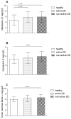
Interleukin-1β (A), interleukin-6 (B), and tumor necrosis factor-α (C) mean serum concentrations in logarithmic scale (±SD) in healthy cows (n = 35) and cows with active (M1, M2, and M4.1; n = 40) or non-active (M3/M4; n = 29) digital dermatitis (DD) lesions. Bonferroni-corrected p-values are presented.
Figure 3
Figure 3
(a) Normal skin (histopathological grade 0, DD M0). The black bar is 0.5 mm in length (HE stain, ×4). (b) Chronic lesion of digital dermatitis without necrosis or inflammation (histopathological grade 4, DD M4). Marked increase in the epidermal thickness by hyperplasia. Some of the dermal papillae are marked with a star and some of the deep epidermal projections are marked with a triangle. The black bar is 0.5 mm in length (HE stain, ×4).
Figure 4
Figure 4
(a) Digital dermatitis, histopathological grade 5 (DD M4). Acute necrosis of the superficial part of the epidermis (HE stain, ×4). (b) Digital dermatitis, histopathological grade 5 (DD M4), same section as in (a). Spirochetes (stained black) are present on the surface of the epidermis and within the necrotic epidermis (black arrows). Viable epidermis is marked with a triangle and necrotic epidermis is marked with a circle (WS stain, ×4).
Figure 5
Figure 5
(a) Digital dermatitis, histopathological grade 5 (DD M4). Acute, locally diffuse necrosis of the superficial part of dermal papillae and the intervening epidermis. Viable epidermis is marked with a triangle, viable dermal papillae with a star, necrotic epidermis with a circle, and necrotic dermal papilla with a square (HE stain, ×4). (b) Digital dermatitis, histopathological grade 5 (DD M4.1). Spirochetes (stained black by silver stain) are present within the necrotic epidermis (black arrows) and in the dermal papillae (white arrows) (WS stain, ×40).
Figure 6
Figure 6
Digital dermatitis, histopathological grade 5 (DD M4.1). Naked, necrotic dermal papillae on the surface of the skin (black arrow) as a result of sloughing off of the necrotic epidermis. Necrotic epidermis is marked with a circle, and necrotic dermal papilla with a square (HE stain, ×10).
Figure 7
Figure 7
Digital dermatitis, histopathological grade 6 (DD M2). Acute ulceration of the skin extending deep into the epidermis and dermis with acute neutrophilic inflammation (area between black lines) (HE stain, ×10).

References

    1. Wilson-Welder J.H., Alt D.P., Nally J.E. Digital Dermatitis in Cattle: Current Bacterial and Immunological Findings. Animals. 2015;5:1114–1135. doi: 10.3390/ani5040400. - DOI - PMC - PubMed
    1. Relun A., Lehebel A., Chesnin A., Guatteo R., Bareille N. Association between digital dermatitis lesions and test-day milk yield of Holstein cows from 41 French dairy farms. J. Dairy Sci. 2013;96:2190–2200. doi: 10.3168/jds.2012-5934. - DOI - PubMed
    1. Mellado M., Saavedra E., Gaytán L., Veliz F.G., Macías-Cruz U., Avendaño-Reyes L., García E. The effect of lameness-causing lesions on milk yield and fertility of primiparous Holstein cows in a hot environment. Livest Sci. 2018;217:8–14. doi: 10.1016/j.livsci.2018.09.008. - DOI
    1. Evans N.J., Murray R.D., Carter S.D. Bovine digital dermatitis: Current concepts from laboratory to farm. Vet. J. 2016;211:3–13. doi: 10.1016/j.tvjl.2015.10.028. - DOI - PubMed
    1. Berry S.L., Read D.H., Famula T.R., Mongini A., Döpfer D. Long-term observations on the dynamics of bovine digital dermatitis lesions on a California dairy after topical treatment with lincomycin HCl. Vet. J. 2012;193:654–658. doi: 10.1016/j.tvjl.2012.06.048. - DOI - PubMed

LinkOut - more resources