. 2024 Jan 31;14(3):462.
doi: 10.3390/ani14030462.

Development of Porcine Accessory Sex Glands

Affiliations

Development of Porcine Accessory Sex Glands

Trish Berger et al. Animals (Basel). .

Abstract

Accessory sex glands are recognized as targets of human disease and may have roles in reproductive success in livestock. The current experiments evaluated the influences of endogenous steroids on the development of porcine accessory sex glands, primarily in the neonatal period. When the aromatase inhibitor, letrozole, was used to inhibit the production of endogenous estrogens in the postnatal interval, growth of the seminal vesicles, prostate, and bulbourethral glands was stimulated. The weights of seminal vesicles, prostate, and bulbourethral glands approximately doubled at 6.5 weeks of age when the reduction in endogenous estrogens began at 1 week of age (p < 0.01). However, by 20 and 40 weeks of age, the weights of accessory sex glands were similar between the letrozole-treated boars and the vehicle-treated littermates indicating the growth stimulation was a transient effect when the treatment interval was short. The presence of both classical nuclear estrogen receptors and the G protein-coupled estrogen receptor in neonatal accessory sex glands indicated multiple signaling pathways might mediate the growth inhibition by endogenous estrogens. The absence of a detectable response when the classical estrogen receptors were blocked with fulvestrant (or when the androgen receptor was blocked with flutamide) suggests that endogenous estrogens act through the G protein-coupled estrogen receptor to inhibit the development of accessory sex glands during this neonatal to early juvenile interval.

Keywords: GPER; bulbourethral glands; estrogen receptor; prostate; seminal vesicles.


Conflict of interest statement

The authors declare no conflicts of interest.

Figures
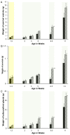
Figure 1
Figure 1
Reducing endogenous estrogens stimulates initial growth of accessory sex glands in pigs. One member of littermate pairs of boars was orally treated weekly with 0.1 mg letrozole/kg bw (hatched bars), and the remaining member was treated with the canola oil vehicle (solid bars). Animals with tissues recovered at 1.3 weeks of age were treated once at 1 day of age; the remaining animals were treated weekly beginning at 1 week of age. Four separate experiments delineated by light background shading consisting of littermate pairs (or littermate sextets for the 2-, 3-, and 5-week experiments) are represented. Values represent least squares means and SEM from the statistical analysis for five animals in each treatment at 1.3, 6.5, and 11 weeks of age and four animals in each age and treatment combination at 2, 3, and 5 weeks of age. * indicates letrozole and vehicle treatments differ with p < 0.05; ** indicates letrozole and vehicle treatments differ with p < 0.01; *** indicates letrozole and vehicle treatments differ with p < 0.0001. (A). Seminal vesicles, (B). prostate, and (C). bulbourethral glands.
Figure 2
Figure 2
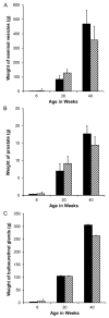
Although reduced endogenous estrogens during the neonatal interval stimulated the initial growth of accessory sex glands and was detectable at 6 weeks of age in these litters (hatched bars represent letrozole treatment), the response did not persist postpuberally when the treatment interval was too short to reprogram aromatase. (A). Seminal vesicles, (B). prostate, and (C). bulbourethral glands. * indicates letrozole and vehicle treatments differ with p < 0.05.
Figure 3
Figure 3
Changes in tissue composition of porcine accessory sex glands during postnatal development. (A). Decrease in relative size of stromal tissue compartment during seminal vesicle development. (B). Increase in relative size of the luminal compartment during seminal vesicle development. (C). Proportion of the seminal vesicle tissue occupied by secretory tissue. (D). Decrease in relative size of stromal tissue compartment during prostate development. (E). Increase in relative size of the luminal compartment during prostate development. (F). Increase in proportion of gland occupied by secretory tissue during prostate development. Values represent the mean of five vehicle-treated boars at 1.3 weeks of age or the least squares mean ± SEM of 3 to 6 vehicle-treated boars with PIC genetic background at older ages. * indicates value is different from preceding time point, p < 0.05; ** indicates value is different from preceding time point, p < 0.01; *** indicates value is different from preceding time point, p < 0.001.
Figure 4
Figure 4
Reducing endogenous estrogens stimulated transitions to more mature tissue composition of accessory sex glands at 11 weeks of age. (A). Proportion of luminal tissue in the seminal vesicles was increased in letrozole-treated littermates (blue bar). (B). Proportion of stromal tissue in the prostate was reduced in letrozole-treated littermates (blue bar). Values are means ± SEM of five littermates. * indicates letrozole and vehicle treatments differ with p < 0.05.
Figure 5
Figure 5
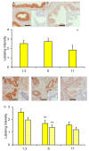
Reduced SOX9 labeling with maturity in the seminal vesicles and prostate. SOX9 labeling in seminal vesicles from 6-week boars (A) and 11-week boars (B). Insets represent tissues incubated with normal serum rather than SOX9 primary antibody. (C). Values represent least squares mean intensity and SEM for glandular epithelium of seminal vesicles for 3 to 6 boars. * indicates a value less than that at the preceding time point, p < 0.05. (D). SOX9 labeling in both the glandular epithelium and the stroma was clearly visible in the prostate from 1.3-week-old boars. (E). Sox9 labeling was less prominent in 1.3-week-old littermates treated with letrozole. (F). Sox9 labeling intensity was less prominent but visible in prostates from 6-week-old boars. Insets represent tissues incubated with normal serum rather than SOX9 antibody. (G). Solid bars represent least squares mean intensity of glandular tissue, and striped bars represent least squares means of stroma tissue of prostate from 3 to 6 boars. ** indicates a value less than that at the preceding time point, p < 0.01. Bars represent 50 microns.
Figure 6
Figure 6
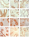
Steroid receptor presence in accessory sex glands from 1.3-week-old boars. Estrogen receptor β (ESR2) immunolabeling (AF), estrogen receptor α (ESR1) immunolabeling (GI), and androgen receptor (AR) immunolabeling (JL) in accessory sex glands from 1.3-week-old boars. Representative sections of seminal vesicle gland from vehicle control boars (A,G,J) and letrozole-treated boars (D) indicate that all three steroid receptors are present and letrozole treatment increased immunolabeling intensity of ESR2 in the glandular epithelium. Representative sections of prostate glands from vehicle control boars (B,H,K) and letrozole-treated boars (E) indicate that all three steroid receptors are present. Representative sections of bulbourethral gland from vehicle control boars (C,I,L) and letrozole-treated boars (F) indicate that all three steroid receptors are present and letrozole-treatment reduced immunolabeling intensity of ESR2 in the bulbourethral gland epithelium. Bar represents 50 µm for all images.
Figure 7
Figure 7
Immunohistochemical localization of GPER in seminal vesicles (A), prostate (B), and bulbourethral glands (C) from 1.3-week-old vehicle-treated boars. Bars represent 25 µm in the seminal vesicles (A) and 100 µm in the prostate (B) and bulbourethral glands (C).
Figure 8
Figure 8
Initial growth of accessory sex glands was stimulated when endogenous estrogens were reduced but not affected if GPER signaling persisted but ESR1 and ESR2 signaling was blocked (fulvestrant). Accessory sex glands were recovered at 6.5 weeks of age and weighed. Values represent least squares means and SEM from the statistical analysis for five animals in each treatment. * indicates values differed from the letrozole treatments with p < 0.05 and ** indicates values differed from the letrozole treatment with p < 0.01.
Figure 9
Figure 9
GPER expression was detectable in the prostate at 2 and 3 weeks of age but expression was not affected by letrozole treatment.

Similar articles

Cited by

References

    1. Izumi K., Mizokami A., Lin W.J., Lai K.P., Chang C. Androgen receptor roles in the development of benign prostate hyperplasia. Am. J. Pathol. 2013;182:1942–1949. doi: 10.1016/j.ajpath.2013.02.028. - DOI - PMC - PubMed
    1. Di Donato M., Giovannelli P., Cernera G., Di Santi A., Marino I., Bilancio A., Galasso G., Auricchio F., Migliaccio A., Castoria G. Non-genomic androgen action regulates proliferative/migratory signaling in stromal cells. Front. Endocrinol. 2014;5:225. doi: 10.3389/fendo.2014.00225. - DOI - PMC - PubMed
    1. Abella D.F., Da Costa M., Guerin Y., Dacheux J.L. Fertility of undiluted ram epididymal spermatozoa stored for several days at 4 °C. Animal. 2015;9:313–319. doi: 10.1017/S1751731114002109. - DOI - PubMed
    1. Berger T., McCarthy M., Pearl C.A., At-Taras E., Roser J.F., Conley A. Reducing endogenous estrogens during the neonatal and juvenile periods affects reproductive tract development and sperm production in postpuberal boars. Anim. Reprod. Sci. 2008;109:218–235. doi: 10.1016/j.anireprosci.2007.10.003. - DOI - PubMed
    1. Rickard J.P., Pini T., Soleilhavoup C., Cognie J., Bathgate R., Lynch G.W., Evans G., Maxwell W.M., Druart X., de Graaf S.P. Seminal plasma aids the survival and cervical transit of epididymal ram spermatozoa. Reproduction. 2014;148:469–478. doi: 10.1530/REP-14-0285. - DOI - PubMed