Review
. 2024 Feb 4;14(3):513.
doi: 10.3390/ani14030513.

Hypothalamic Neuromodulation of Hypothermia in Domestic Animals

Affiliations
Review

Hypothalamic Neuromodulation of Hypothermia in Domestic Animals

Daniel Mota-Rojas et al. Animals (Basel). .

Abstract

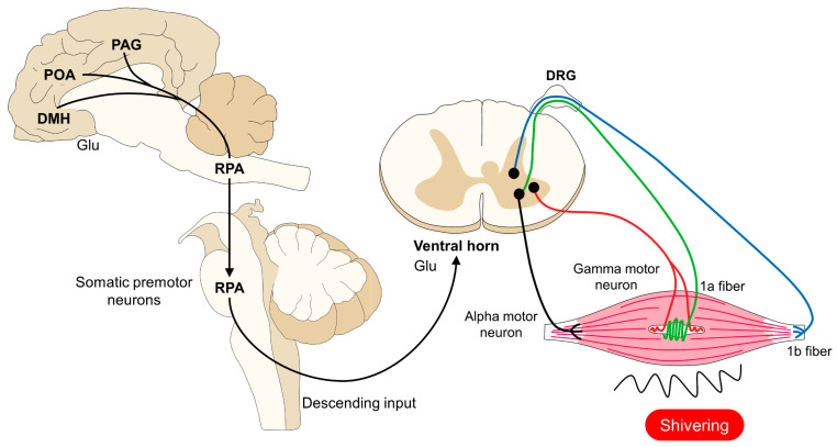
When an organism detects decreases in their core body temperature, the hypothalamus, the main thermoregulatory center, triggers compensatory responses. These responses include vasomotor changes to prevent heat loss and physiological mechanisms (e.g., shivering and non-shivering thermogenesis) for heat production. Both types of changes require the participation of peripheral thermoreceptors, afferent signaling to the spinal cord and hypothalamus, and efferent pathways to motor and/or sympathetic neurons. The present review aims to analyze the scientific evidence of the hypothalamic control of hypothermia and the central and peripheral changes that are triggered in domestic animals.

Keywords: brown adipose tissue thermogenesis; cold-defensive behaviors; cutaneous vasoconstriction; infrared thermography.


Conflict of interest statement

The authors declare no conflicts of interest.

Figures

Figure 1
Figure 1
Hypothalamic control of hypothermia in domestic animals. The perception of low core body temperatures is due to the presence of thermoreceptors (TRPM8 and TRPA1). These receptors project to the DRG and DH in the spinal cord, where glutamatergic neurons transmit the thermal information to the POA. To generate thermogenic responses, connections from the POA to the PO, DMH, and rMR activate sympathetic or motor neurons that, in consequence, cause cutaneous vasoconstriction, BAT thermogenesis, or skeletal muscle shivering. Ach: acetylcholine; BAT: brown adipose tissue; Glu: glutamate; DH: dorsal horn; DMH: dorsomedial hypothalamus; DRG: dorsal root ganglion; IML: intermediolateral laminae; LPBel: lateral parabrachial nucleus lateral part; MnPO: median preoptic area; MPO: medial preoptic area; NA: noradrenaline; POA: preoptic area; RPa: rostral raphe pallidus; and 5-HT: serotonin.
Figure 2
Figure 2
Modulation of cutaneous vasoconstriction and BAT thermogenesis. When the POA detects a decrease in core body temperature, two main responses are triggered. On the one hand, the neural pathway (marked with a non-continuous line) activates sympathetic CVC premotor neurons in the RPA to send glutamatergic and serotoninergic inputs to the IML. From these neurons, sympathetic axons innervating the endothelium of dermal blood vessels respond to NA release, eliciting vasoconstriction. On the other hand, the activation of premotor neurons innervating the BAT cause thermogenesis by the lipolysis of the BAT. ACh: acetylcholine; BAT: brown adipose tissue; DMH: dorsomedial hypothalamus; Glu: glutamate; IML: intermediolateral laminae; LH: lateral hypothalamus; NA: noradrenaline; PAG: periaqueductal gray; POA: preoptic area; RPA: rostral raphe pallidus; VTA: ventral tegmental area; and 5-HT: serotonin.
Figure 3
Figure 3
Thermal responses associated with the administration of general anesthetics. (A). The surface temperature of a male European domestic cat before anesthetic management for an advanced imaging diagnostic procedure is shown. The surface temperature at the periocular level (El1) recorded a maximum temperature of 36.2 °C, an average of 35.6 °C, and a minimum of 34.7 °C. (B). After the induction of anesthesia with propofol and maintenance with isoflurane, the temperature at the average periocular temperature (El1) decreased up to 3.6 °C. The decrease in surface temperature is due to the vasodilation that the anesthetic causes, allowing the redistribution of temperature. The maximum temperature is indicated with a red triangle, and the minimum temperature is indicated with a blue triangle.
Figure 4
Figure 4
Differences between the surface temperatures of the peripheral and central regions in newborn animals. The differences in the surface temperature found between the central region (periocular, El1) and the peripheral region (thoracic or pelvic limb, El2) in both water buffaloes (A) and newborn piglets (B) are shown. In the case of the newborn water buffalo, an average difference in the temperature between both regions was set at 4.1 °C. In the piglet, an average difference in the maximum temperature of both regions was set at 9.4 °C. The possible explanation for this fact is that, during the perception of cold, the sympathetic postganglionic fibers release adrenaline and norepinephrine, which act on the α-2 adrenergic receptors present in the endothelium. This causes the vasoconstriction of the peripheral blood vessels that reduce the level of heat loss, preserving the temperature in central areas such as the head and thorax. The maximum temperature is indicated with a red triangle, and the minimum temperature is indicated with a blue triangle. Radiometric images were obtained using a T1020 FLIR thermal camera. Image resolution: 1024 × 768; up to 3.1 MP with UltraMax. FLIR Systems, Inc. Wilsonville, OR, USA.
Figure 5
Figure 5
Comparison of surface temperatures in a newborn dog. (A) Central thermal windows. The upper (Li1) and lower (Li2) eyelids marked by a line are the central windows that are used to evaluate the thermal states of newborn puppies. The maximum temperature of these regions is 35.1 °C. In contrast, the (B) thoracic limb metacarpal (El1) and elbow (El2) thermal windows show lower temperatures by up to 0.8 °C. The differences in temperatures can be attributed to the immature thermoregulatory mechanisms in newborn dogs, which forces the organism to elicit changes to prevent heat loss (peripheral vasoconstriction) and produce heat (motivation to consume colostrum). The maximum temperature is indicated with a red triangle, and the minimum temperature is indicated with a blue triangle.
Figure 6
Figure 6
Central and peripheral control of shivering thermogenesis. Similar to the vasomotor and BAT thermogenesis, the POA, DMH, PAG, and RPA modulate muscular contractions. In the RPA, premotor neurons respond to hypothermia by activating alpha and gamma neurons that directly innervate muscle fibers. The sustained muscular contraction produces heat to promote an increase in core body temperature. ACh: acetylcholine; DMH: dorsomedial hypothalamus; DRG: dorsal root ganglion; Glu: glutamate; IML: intermediolateral laminae; PAG: periaqueductal gray; POA: preoptic area; and RPA: rostral raphe pallidus.
Figure 7
Figure 7
The importance of thermoregulatory behaviors and neonatal development in newborn rat pups. (A) The influence of huddling as a thermoregulatory behavior highly influences the thermal states of newborn rats during the first weeks of life. It can be observed that the maximum interscapular temperature of the pup in the center of the nest (El1) is 0.6 °C higher than the values recorded in a pup located in the nest periphery (El2). (B) shows the thermoregulatory advantage that hair growth has on the surface temperature of animals. In a 12-day-old rat pup with fur growth, the maximum temperature at the interscapular space (El1) is 35.6 °C. In contrast, in newborn pups (C), the maximum temperature of the same region is 0.4 °C lower, although both animals are inside the nest with their conspecifics. These thermal images help us to understand that behavioral adjustments play an essential role in mammal thermoregulation. The maximum temperature is indicated with a red triangle, and the minimum temperature is indicated with a blue triangle. Radiometric images were obtained using a T1020 FLIR thermal camera. Image resolution: 1024 × 768; up to 3.1 MP with UltraMax. FLIR Systems, Inc. Wilsonville, OR, USA.

References

    1. Osilla E., Marsidi J., Sharma S. Physiology, Temperature Regulation. StatPearls Publishing; Tampa, FL, USA: 2018. - PubMed
    1. Collier R.J., Gebremedhin K.G. Thermal Biology of Domestic Animals. Annu. Rev. Anim. Biosci. 2015;3:513–532. doi: 10.1146/annurev-animal-022114-110659. - DOI - PubMed
    1. Krishnan G., Silpa M.V., Sejian V. Textbook of Veterinary Physiology. Springer Nature Singapore; Singapore: 2023. Environmental Physiology and Thermoregulation in Farm Animals; pp. 723–749.
    1. Oka T. Handbook of Clinical Neurology. Volume 157. Elsevier; Amsterdam, The Netherlands: 2018. Stress-Induced Hyperthermia and Hypothermia; pp. 599–621. - PubMed
    1. Erecinska M., Thoresen M., Silver I.A. Effects of Hypothermia on Energy Metabolism in Mammalian Central Nervous System. J. Cereb. Blood Flow Metab. 2003;23:513–530. doi: 10.1097/01.WCB.0000066287.21705.21. - DOI - PubMed

LinkOut - more resources