

Early Biofilm Formation on the Drain Tip after Total Knee Arthroplasty Is Not Associated with Prosthetic Joint Infection: A Pilot Prospective Case Series Study of a Single Center
- PMID: 38338251
- PMCID: PMC10855896
- DOI: 10.3390/healthcare12030366
Early Biofilm Formation on the Drain Tip after Total Knee Arthroplasty Is Not Associated with Prosthetic Joint Infection: A Pilot Prospective Case Series Study of a Single Center
Abstract
Background: Periprosthetic joint infection (PJI) is a devastating complication of arthroplasties that could occur during the surgery. The purpose of this study was to analyze the biofilm formation through microbiological culture tests and scanning electron microscopy (SEM) on the tip of surgical drainage removed 24 h after arthroplasty surgery.
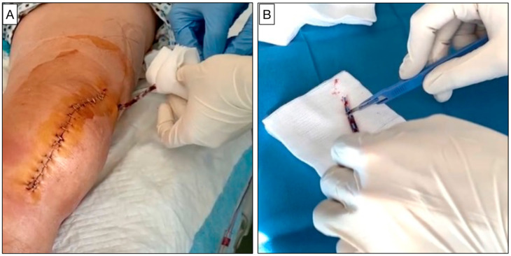
Methods: A total of 50 consecutive patients were included in the present prospective observational study. Drains were removed under total aseptic conditions twenty-four hours after surgery. The drain tip was cut in three equal parts of approximately 2-3 cm in length and sent for culture, culture after sonication, and SEM analysis. The degree of biofilm formation was determined using a SEM semi-quantitative scale.
Results: From the microbiological analysis, the cultures of four samples were positive. The semi-quantitative SEM analysis showed that no patient had grade 4 of biofilm formation. A total of 8 patients (16%) had grade 3, and 14 patients (28%) had grade 2. Grade 1, scattered cocci with immature biofilm, was contemplated in 16 patients (32%). Finally, 12 patients (24%) had grade 0 with a total absence of bacteria. During the follow-up (up to 36 months), no patient showed short- or long-term infectious complications.
Conclusions: Most of the patients who underwent primary total knee arthroplasty (TKA) showed biofilm formation on the tip of surgical drain 24 h after surgery even though none showed a mature biofilm formation (grade 4). Furthermore, 8% of patients were characterized by a positivity of culture analysis. However, none of the patients included in the study showed signs of PJI up to 3 years of follow-up.
Keywords: biofilm; infection disease; periprosthetic joint infection; surgical drainage; total knee arthroplasty.
Conflict of interest statement
The authors declare no conflicts of interest.
Figures


Similar articles
-
Drain tip cultures do not predict infections in primary total knee arthroplasty.Clin Ter. 2015;166(3):e153-7. doi: 10.7417/CT.2015.1846. Clin Ter. 2015. PMID: 26152624
-
The Function of Sonication in the Diagnosis of Periprosthetic Joint Infection After Total Knee Arthroplasty.Arch Bone Jt Surg. 2022 Sep;10(9):735-740. doi: 10.22038/abjs.2020.44329.2212. Arch Bone Jt Surg. 2022. PMID: 36246019 Free PMC article. Review.
-
Superiority of the sonication method against conventional periprosthetic tissue cultures for diagnosis of prosthetic joint infections.Eur J Orthop Surg Traumatol. 2018 Jan;28(1):51-57. doi: 10.1007/s00590-017-2012-y. Epub 2017 Jul 17. Eur J Orthop Surg Traumatol. 2018. PMID: 28714050
-
Intrawound vancomycin powder increases post-operative wound complications and does not decrease periprosthetic joint infection in primary total and unicompartmental knee arthroplasties.Knee Surg Sports Traumatol Arthrosc. 2019 Jul;27(7):2322-2327. doi: 10.1007/s00167-019-05498-z. Epub 2019 Apr 9. Knee Surg Sports Traumatol Arthrosc. 2019. PMID: 30968239
-
Sonication of Arthroplasty Implants Improves Accuracy of Periprosthetic Joint Infection Cultures.Clin Orthop Relat Res. 2017 Jul;475(7):1827-1836. doi: 10.1007/s11999-017-5315-8. Clin Orthop Relat Res. 2017. PMID: 28290115 Free PMC article. Review.
References
LinkOut - more resources
Full Text Sources
