. 2024 Jan 24;25(3):1443.
doi: 10.3390/ijms25031443.

Inhibitory Effect of a Tankyrase Inhibitor on Mechanical Stress-Induced Protease Expression in Human Articular Chondrocytes

Affiliations

Inhibitory Effect of a Tankyrase Inhibitor on Mechanical Stress-Induced Protease Expression in Human Articular Chondrocytes

Yoshifumi Hotta et al. Int J Mol Sci. .

Abstract

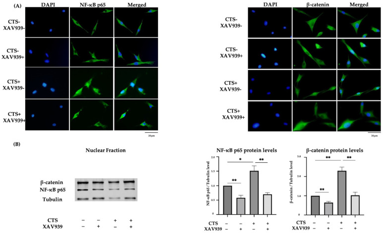
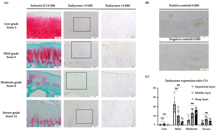
We investigated the effects of a Tankyrase (TNKS-1/2) inhibitor on mechanical stress-induced gene expression in human chondrocytes and examined TNKS-1/2 expression in human osteoarthritis (OA) cartilage. Cells were seeded onto stretch chambers and incubated with or without a TNKS-1/2 inhibitor (XAV939) for 12 h. Uni-axial cyclic tensile strain (CTS) (0.5 Hz, 8% elongation, 30 min) was applied and the gene expression of type II collagen a1 chain (COL2A1), aggrecan (ACAN), SRY-box9 (SOX9), TNKS-1/2, a disintegrin and metalloproteinase with thrombospondin motifs-5 (ADAMTS-5), and matrix metalloproteinase-13 (MMP-13) were examined by real-time PCR. The expression of ADAMTS-5, MMP-13, nuclear translocation of nuclear factor-κB (NF-κB), and β-catenin were examined by immunocytochemistry and Western blotting. The concentration of IL-1β in the supernatant was examined by enzyme-linked immunosorbent assay (ELISA). TNKS-1/2 expression was assessed by immunohistochemistry in human OA cartilage obtained at the total knee arthroplasty. TNKS-1/2 expression was increased after CTS. The expression of anabolic factors were decreased by CTS, however, these declines were abrogated by XAV939. XAV939 suppressed the CTS-induced expression of catabolic factors, the release of IL-1β, as well as the nuclear translocation of NF-κB and β-catenin. TNKS-1/2 expression increased in mild and moderate OA cartilage. Our results demonstrated that XAV939 suppressed mechanical stress-induced expression of catabolic proteases by the inhibition of NF-κB and activation of β-catenin, indicating that TNKS-1/2 expression might be associated with OA pathogenesis.

Keywords: ADAMTS-5; IL-1β; MMP-13; NF-κB; SOX9; XAV939; chondrocyte; mechanical stress; osteoarthritis; tankyrases; β-catenin.


Conflict of interest statement

The authors declare no conflicts of interest.

Figures

Figure 1
Figure 1
(A) Expression of anabolic factors after cyclic tensile strain in normal human chondrocytes. The expression of each factor decreased up to 12 h after the application of CTS. (B) Expression of catabolic factors after CTS. The peak expression was at 12 h after CTS. (C) The expression of TNKS-1/2 after CTS. The peak expression was at 12 h after CTS. (D) The increase in TNKS-1/2 protein levels after CTS by Western blot analysis and quantification of TNKS-1/2 protein levels (* p < 0.05, ** p < 0.01, ns: not significant).
Figure 2
Figure 2
(A) Relative expression of CTNNB1 mRNA in normal human chondrocyte cells, treated with different XAV939 concentrations (0, 1, 10, and 100 μM). (BD) The effects of a TNKS-1/2 inhibitor (XAV939: 10 μM) on the expression of anabolic (B), and catabolic factors (C) and TNKS-1/2 (D) in human chondrocytes at 12 h after CTS. (E) The concentration of IL-1β in culture medium after CTS with or without XAV939 (* p < 0.05, ** p < 0.01, *** p < 0.0001, ns: not significant).
Figure 3
Figure 3
Western blots for total ERK and phosphorylated ERK (p-ERK) (A), total p38 and phosphorylated p38 (p-p38) (B), or total JNK and phosphorylated JNK (p-JNK) (C) in normal human chondrocytes (* p < 0.05, ** p < 0.01, ns: not significant).
Figure 4
Figure 4
(A) The effect of mechanical stress and XAV939 on the expression of ADAMTS-5 and MMP-13 in normal human chondrocytes. Up-regulation of ADAMTS-5 and MMP-13 was localized to the cytoplasm (DAPI: blue signal, ADAMTS-5 and MMP-13: green signal). Scale bar = 20 μm. (B) The percentages of chondrocytes positive for ADAMTS-5 and MMP-13. Cell numbers were counted in four fields, at 10× magnification, and the mean was calculated (* p < 0.05, ** p < 0.01, *** p < 0.0001). (C) The effect of XAV939 on the protein expression of ADAMTS-5 and MMP-13 by Western blot and quantification analysis.
Figure 5
Figure 5
(A) The effect of mechanical stress and XAV939 on nuclear translocation of NF-κB p65 and β-catenin by immunocytochemistry in normal human chondrocytes (DAPI: blue signal, NF-κB p65 and β-catenin: green signal). Scale bar = 20 μm. (B) Western blot analysis and quantification of nuclear extraction proteins for NF-κB p65 and β-catenin. (* p < 0.05, ** p < 0.01).
Figure 6
Figure 6
(A) Double immunohistochemical staining of β-catenin and NF-κB p65 nuclear translocation in normal human chondrocytes (DAPI: blue signal, β-catenin: green signal, NF-κB p65: red signal). Arrows showed double staining cells. (B) Both β-catenin and NF-κB p65 translocation to nuclei were observed after CTS without XAV939, while this effect was canceled by XAV939 (** p < 0.01).
Figure 7
Figure 7
Immunohistochemical evaluation of TNKS-1/2 in human articular cartilage tissues. (A) In tissues with low-grade OA (Mankin score 0–2), TNKS-1/2 expression was not clear. In tissue with mild-grade OA (Mankin score 3–6), the main expression of TNKS-1/2 was in superficial and deep layers. In tissue with moderate-grade OA (Mankin score 7–10), the main expression of TNKS-1/2 was in the middle to deep layers. In tissue with severe-grade OA (Mankin score 11–14), TNKS-1/2 expression was observed in the deep layer but not in the superficial and middle layers. (B) No signal was observed in the negative control. (C) In low-grade OA, TNKS-1/2 were rarely expressed in every layer. In mild-grade OA, TNKS-1/2 expression was significantly increased in the superficial layer relative to the middle and deep layers. In moderate-grade OA, expression was significantly increased in the deep and middle layers compared to the superficial layer. In severe-grade OA, the expression was low relative to other grades and the difference between layers was not clear (** p < 0.01, ns: not significant). The scale bars were set at 200 μm at 100× magnification and 100 μm at 200× magnification.

References

    1. Dequeker J., Luyten F.P. The history of osteoarthritis-osteoarthrosis. Ann. Rheum. Dis. 2008;67:5–10. doi: 10.1136/ard.2007.079764. - DOI - PubMed
    1. Loeser R.F. The Role of Aging in the Development of Osteoarthritis. Trans. Am. Clin. Climatol. Assoc. 2017;128:44–54. - PMC - PubMed
    1. Yao Q., Wu X., Tao C., Gong W., Chen M., Qu M., Zhong Y., He T., Chen S., Xiao G. Osteoarthritis: Pathogenic signaling pathways and therapeutic targets. Signal Transduct. Target. Ther. 2023;8:56. doi: 10.1038/s41392-023-01330-w. - DOI - PMC - PubMed
    1. Loeser R.F., Goldring S.R., Scanzello C.R., Goldring M.B. Osteoarthritis: A disease of the joint as an organ. Arthritis Rheum. 2012;64:1697–1707. doi: 10.1002/art.34453. - DOI - PMC - PubMed
    1. Ushiyama T., Chano T., Inoue K., Matsusue Y. Cytokine production in the infrapatellar fat pad: Another source of cytokines in knee synovial fluids. Ann. Rheum. Dis. 2003;62:108–112. doi: 10.1136/ard.62.2.108. - DOI - PMC - PubMed

MeSH terms