. 2024 Jan 30;25(3):1669.
doi: 10.3390/ijms25031669.

Extended Cleavage Specificity of two Hematopoietic Serine Proteases from a Ray-Finned Fish, the Spotted Gar (Lepisosteus oculatus)

Affiliations

Extended Cleavage Specificity of two Hematopoietic Serine Proteases from a Ray-Finned Fish, the Spotted Gar (Lepisosteus oculatus)

Paolo Valentini et al. Int J Mol Sci. .

Abstract

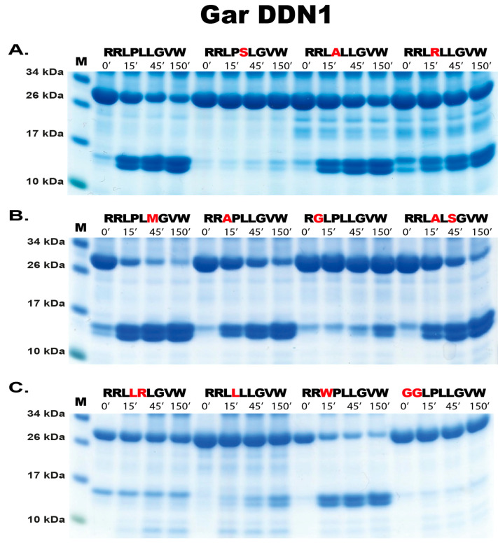
The extended cleavage specificities of two hematopoietic serine proteases originating from the ray-finned fish, the spotted gar (Lepisosteus oculatus), have been characterized using substrate phage display. The preference for particular amino acids at and surrounding the cleavage site was further validated using a panel of recombinant substrates. For one of the enzymes, the gar granzyme G, a strict preference for the aromatic amino acid Tyr was observed at the cleavable P1 position. Using a set of recombinant substrates showed that the gar granzyme G had a high selectivity for Tyr but a lower activity for cleaving after Phe but not after Trp. Instead, the second enzyme, gar DDN1, showed a high preference for Leu in the P1 position of substrates. This latter enzyme also showed a high preference for Pro in the P2 position and Arg in both P4 and P5 positions. The selectivity for the two Arg residues in positions P4 and P5 suggests a highly specific substrate selectivity of this enzyme. The screening of the gar proteome with the consensus sequences obtained by substrate phage display for these two proteases resulted in a very diverse set of potential targets. Due to this diversity, a clear candidate for a specific immune function of these two enzymes cannot yet be identified. Antisera developed against the recombinant gar enzymes were used to study their tissue distribution. Tissue sections from juvenile fish showed the expression of both proteases in cells in Peyer's patch-like structures in the intestinal region, indicating they may be expressed in T or NK cells. However, due to the lack of antibodies to specific surface markers in the gar, it has not been possible to specify the exact cellular origin. A marked difference in abundance was observed for the two proteases where gar DDN1 was expressed at higher levels than gar granzyme G. However, both appear to be expressed in the same or similar cells, having a lymphocyte-like appearance.

Keywords: cleavage specificity; evolution; fish; macrophage; serine protease; tryptase.


Conflict of interest statement

The authors declare no conflict of interest.

Figures

Figure 1
Figure 1
Nomenclature of the amino acids surrounding the cleavage site and the amino acids forming the active site pocket. In panel (A), the nomenclature of the amino acids surrounding the cleaved peptide bond is shown. The amino acid N-terminals from the cleaved bond are termed as P1 (where cleavage occurs, depicted by scissors), P2, P3, etc. Amino acid C-terminals of the cleaved bond are termed as P1′ (adjacent to P1), P2′, P3′, etc. The corresponding interacting sub-sites in the enzyme are denoted with S. In panel (B), the three amino acids forming the active site pocket (S1 pocket) are shown. These three residues correspond to positions 189, 216, and 226 in bovine pancreatic chymotrypsinogen and have been found to determine the primary specificity of the enzyme as either chymotrypsin-, trypsin-, or elastase-like specificity [31]. The preferred amino acids in the P1 position of the corresponding substrates are illustrated in green, yellow, and blue, respectively. Figure reproduced from [32].
Figure 2
Figure 2
Phylogenetic relationship between gar granzyme G, gar DDN1, and other hematopoietic serine proteases. All sequences were run in the multiple alignment programme, MAFFT, to verify if they belonged to the serine protease family. The tree was constructed using MRBAYES with a Bayesian interference of phylogeny algorithm (with posterior probabilities), opened with FigTree (v1.4) and annotated in Adobe illustrator (CS5). Panel (A) shows the entire analysis involving a total of 368 vertebrate serine protease sequences. Panel (B) shows an enlargement of the branch of the major tree, where the majority of the fish proteases are found, except the granzyme A/K-related fish proteases. The proteases of particular interest for this study are marked with red arrows. All genes for proteins that were produced as recombinant proteins are marked with red stars. Figure adapted from [32].
Figure 3
Figure 3
Recombinant gar granzyme G and gar DDN1. The enzymes were produced as an inactive protein (left lane) in HEK293 cells with an N-terminal His6-tag and enterokinase (EK) site, facilitating purification and activation, respectively. The addition of EK cleaves the N-terminal sites, resulting in an active enzyme and a subsequent drop in size (right lane). The enzymes were run on a 4–12% pre-cast SDS-PAGE gel (Invitrogen, Carlsbad, CA, USA) and stained with colloidal Coomassie brilliant blue.
Figure 4
Figure 4
Substrate phage display sequences. The phage display-derived sequences from the analysis of both gar granzyme G and gar DDN1 are displayed in the left part of the figure. Each line represents a separate phage-derived sequence. For comparison, two previous phage display analyses are included, with specificities similar to what was observed for gar granzyme G and gar DDN1. Gar granzyme G, which has a P1 preference for Tyr, shows similarities to a recently analyzed Zebra mbuna enzyme named granzyme A2 [34]. The gar DDN1 shows some similarities to rabbit and guinea pig Leu-ases, as they share the same P1 preference [35].
Figure 5
Figure 5
Verification of phage display sequences using the 2xTrx system. A number of phage display-derived sequences and variants of these sequences were added in between two adjacent trx proteins (panel (A), adapted from [32]), expressed in E. coli and subjected to gar granzyme G (panels (C,D)). The results were run on pre-cast 4–12% SDS-PAGE gels (Invitrogen, Carlsbad, CA, USA). Hypothetical cleavage is shown (panel (B)) to highlight the possible cleavage patterns. The individual lanes represent various time points after the addition of the enzyme, in minutes.
Figure 6
Figure 6
Verification of phage display sequences of gar DDN1 using the 2xTrx system. A number of phage display-derived sequences and variants of these sequences were added in between two adjacent trx proteins, were expressed in E. coli, and were subjected to gar DDN1 (panels (AC)). The results were run on pre-cast 4–12% SDS-PAGE gels (Invitrogen, Carlsbad, CA, USA). The individual lanes represent various time points after the addition of the enzyme, in minutes.
Figure 7
Figure 7
Histological analysis of the gar tissues. Panel (A) shows a picture of a gar. Panel (B) shows a picture of the juvenile gar used for the histological analysis of head kidney and intestinal regions for immune cells. The section of the animal that was used for analysis is marked with a white line and a red arrowhead. Panels (CF) show hematoxylin–eosin-stained tissue sections. Panel (C) shows primarily the intestinal region with a white arrowhead (1) pointing at a Peyer’s patch-like structure in the intestinal wall. Panel (D) shows an enlargement of this Peyer’s patch region (arrowhead 2). Panels (E,F) show the head kidney region with arrowheads, marking the pure red-blood-cell-rich regions of the head kidney (arrowhead 3). Arrowhead V shows the vertebra. In panel (E), arrowhead number 4 shows an enlargement of this red blood region, and arrowhead 5 shows the immune-cell-rich region of the head kidney.
Figure 8
Figure 8
Staining using DDN1 and GZM-G antibodies. In panel (A), a representative section of DDN1 staining is shown, and in (B), a magnification of this section is shown for clarity. In panel (C), a representative section of the staining of gzm-G antibody is shown, and in panel (D), the primary antibody control of fish tissue is shown. Nuclei were stained using DAPI in blue.
Figure 9
Figure 9
Genomic loci encoding fish hematopoietic serine proteases. The locus encoding both enzymes is encoded in the ray-finned fish met-ase locus. This locus also exists in mammals, including humans, with a slightly different organization. The human locus has most likely experienced an inversion and an internal expansion, marked with a yellow oval just below the human locus. Serine protease genes have a double height for visual identification. Red dots mark the two enzymes analyzed in this communication, and blue dots mark enzymes that were previously analyzed. Green dots mark the two catfish proteases analyzed in [32] and the two zebra fish proteases that show similarities to the cafish proteases discussed in [32] and in the discussion of this manuscript. Panel (A) shows a tree of fish evolution based on both genetic and morphological information, where bichir is the first to branch out from the common fish branch followed by starlet and later the gar and finally the teleosts [24,36]. Panel (B) shows the loci of the different fish species specified in panel (A).

References

    1. Hellman L., Thorpe M. Granule proteases of hematopoietic cells, a family of versatile inflammatory mediators—An update on their cleavage specificity, in vivo substrates, and evolution. Biol. Chem. 2014;395:15–49. doi: 10.1515/hsz-2013-0211. - DOI - PubMed
    1. Akula S., Thorpe M., Boinapally V., Hellman L. Granule Associated Serine Proteases of Hematopoietic Cells—An Analysis of Their Appearance and Diversification during Vertebrate Evolution. PLoS ONE. 2015;10:e0143091. doi: 10.1371/journal.pone.0143091. - DOI - PMC - PubMed
    1. Fu Z., Thorpe M., Akula S., Chahal G., Hellman L. Extended cleavage specificity of human neutrophil elastase, human proteinase 3 and their distant orthologue clawed frog PR3-three elastases with similar primary but different extended specificities and stability. Front. Immunol. 2018;9:2387. doi: 10.3389/fimmu.2018.02387. - DOI - PMC - PubMed
    1. Fu Z., Thorpe M., Alemayehu R., Roy A., Kervinen J., de Garavilla L., Abrink M., Hellman L. Highly Selective Cleavage of Cytokines and Chemokines by the Human Mast Cell Chymase and Neutrophil Cathepsin G. J. Immunol. 2017;198:1474–1483. doi: 10.4049/jimmunol.1601223. - DOI - PubMed
    1. Akula S., Fu Z., Wernersson S., Hellman L. The Evolutionary History of the Chymase Locus -a Locus Encoding Several of the Major Hematopoietic Serine Proteases. Int. J. Mol. Sci. 2021;22:10975. doi: 10.3390/ijms222010975. - DOI - PMC - PubMed

LinkOut - more resources