. 2024 Jan 30;25(3):1696.
doi: 10.3390/ijms25031696.

A Blood Test for the Diagnosis of Multiple Sclerosis

Affiliations

A Blood Test for the Diagnosis of Multiple Sclerosis

Paola Giuliano et al. Int J Mol Sci. .

Abstract

Multiple sclerosis (MS) is an autoimmune chronic disease characterized by inflammation and demyelination of the central nervous system (CNS). Despite numerous studies conducted, valid biomarkers enabling a definitive diagnosis of MS are not yet available. The aim of our study was to identify a marker from a blood sample to ease the diagnosis of MS. In this study, since there is evidence connecting the serotonin pathway to MS, we used an ELISA (Enzyme-Linked Immunosorbent Assay) to detect serum MS-specific auto-antibodies (auto-Ab) against the extracellular loop 1 (ECL-1) of the 5-hydroxytryptamine (5-HT) receptor subtype 2A (5-HT2A). We utilized an ELISA format employing poly-D-lysine as a pre-coating agent. The binding of 208 serum samples from controls, both healthy and pathological, and of 104 serum samples from relapsing-remitting MS (RRMS) patients was tested. We observed that the serum-binding activity in control cohort sera, including those with autoimmune and neurological diseases, was ten times lower compared to the RRMS patient cohort (p = 1.2 × 10-47), with a sensitivity and a specificity of 98% and 100%, respectively. These results show that in the serum of patients with MS there are auto-Ab against the serotonin receptor type 2A which can be successfully used in the diagnosis of MS due to their high sensitivity and specificity.

Keywords: 5-HT2A receptor; ELISA assay; blood serum; diagnosis; multiple sclerosis; peptides.


Conflict of interest statement

R.P. has received research grants from PRINDEX S.r.l.; P.G., M.S. and R.P are equity owners in PRINDEX S.r.l.; P.G., G.L.R., S.C. and S.D. have been involved as consultants in PRINDEX S.r.l.; L.M.P., M.S. and R.P. are the inventors of USA patent 10633427 and EPO patent 3109257.

Figures

Figure 1
Figure 1
Comparison between coating in a carbonate/bicarbonate buffer versus poly-D-lysine. Dose-response curves on the two ELISA formats, performed with coating of a carbonate/bicarbonate buffer pH 9.6 (A) or with a previous pre-coat with 10 μg/mL of poly-D-lysine (B). The results are the mean ± SEM of n = 12 to 24 CTRL and RRMS sera performed in 3 independent experiments. The results are expressed as OD450 minus background. p-values were calculated with ANOVA (C). Intra-assay comparison of both ELISA formats on a panel of 10 CTRL and 8 RRMS sera in 3 independent experiments. The results are expressed as S/P values, as specified in Materials and Methods. p-values (calculated using the Mann–Whitney t-test) of significant differences are reported above the plot as asterisks. The asterisks ** and *** indicates, respectively, p ≤ 0.01 and ≤0.001.
Figure 1
Figure 1
Comparison between coating in a carbonate/bicarbonate buffer versus poly-D-lysine. Dose-response curves on the two ELISA formats, performed with coating of a carbonate/bicarbonate buffer pH 9.6 (A) or with a previous pre-coat with 10 μg/mL of poly-D-lysine (B). The results are the mean ± SEM of n = 12 to 24 CTRL and RRMS sera performed in 3 independent experiments. The results are expressed as OD450 minus background. p-values were calculated with ANOVA (C). Intra-assay comparison of both ELISA formats on a panel of 10 CTRL and 8 RRMS sera in 3 independent experiments. The results are expressed as S/P values, as specified in Materials and Methods. p-values (calculated using the Mann–Whitney t-test) of significant differences are reported above the plot as asterisks. The asterisks ** and *** indicates, respectively, p ≤ 0.01 and ≤0.001.
Figure 2
Figure 2
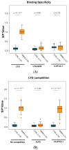
Analytical specificity on the poly-D-lysine ELISA format. Binding specificity on the poly-D-lysine ELISA format was assayed by (A) cross-reactivity on control peptides and (B) by competition experiments. (A) RRMS sera show binding activity only on the LYG peptide, whilst control peptides (LYG-SCR and 5-HT1A-1) did not react with RRMS sera. (B) Competition experiments were conducted by pre-incubation of sera with 5 μM of specific (+LYG) or non-specific (+5HT2A-1) peptide before plating on LYG-coated wells. The results are expressed as S/P values, as specified in Materials and Methods. More than 3 independent experiments were performed for each assay. All peptides were coated at 25 μM. RRMS and controls were compared using the Mann–Whitney t-test, and the p-values are reported above each plot.
Figure 3
Figure 3
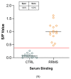
Serum compounds do not interfere with auto-Ab binding. IgG purified (10 μM) from a panel of 15 RRMS and 15 control samples was assayed in parallel with serum specimens (1:100 dilution) in 3 independent experiments. RRMS sera (A) and IgG (B) showed comparable binding activity on LYG. A horizontal red line indicated the cut-off value of 0.40 yielding the best sensitivity and specificity. RRMS and controls were compared using the Mann–Whitney t-test, and the p-values are reported above plot (C).
Figure 3
Figure 3
Serum compounds do not interfere with auto-Ab binding. IgG purified (10 μM) from a panel of 15 RRMS and 15 control samples was assayed in parallel with serum specimens (1:100 dilution) in 3 independent experiments. RRMS sera (A) and IgG (B) showed comparable binding activity on LYG. A horizontal red line indicated the cut-off value of 0.40 yielding the best sensitivity and specificity. RRMS and controls were compared using the Mann–Whitney t-test, and the p-values are reported above plot (C).
Figure 4
Figure 4
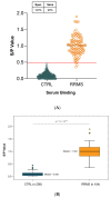
Serum autoantibody reactivity against LYG peptide. An ELISA assay was used to detect anti-5-HT2A serum auto-Ab. LYG peptide was coated on poly-D-lysine pre-coated microplates (A) The dot plot represents S/P values in 208 controls and 104 RRMS cases. A horizontal red line indicated the cut-off value of 0.48 yielding the best sensitivity and specificity (98% and 100%, respectively). (B) Box-and-whisker plots showing the distribution of S/P values in RRMS (median 1.004; IQR 0.814–1.172) and CTRL (median 0.084; IQR 0.040–0.143). RRMS and CTRL samples were compared using the Mann–Whitney t-test, and the p-values are reported above the plot.
Figure 5
Figure 5
Autoimmune/neurological diseases and Disease-Modifying Treatment (DMT) do not affect LYG binding: the same data as used in Figure 4 are represented in the subgroups that made up each MS and control population. The statistical comparison between the subgroups (using the Kruskal–Wallis rank–sum test) revealed no significant differences within each cohort (control p = 0.051 for 10 groups and MS p = 0.779 for 8 groups). CTRL: healthy controls; ND: Neurologic Diseases; CIDP: Chronic Inflammatory Demyelinating Polyneuropathy; MG: Myasthenia Gravis; MH: Migraine Headache; AR: Rheumatoid Arthritis; LES: Lupus Erythematosus; SJS: Sjogren; SS: Systemic Sclerosis; CD: Coeliachia Disease.

References

    1. Brownlee W.J., Hardy T.A., Fazekas F., Miller D.H. Diagnosis of multiple sclerosis: Progress and challenges. Lancet. 2017;389:1336–1346. doi: 10.1016/S0140-6736(16)30959-X. - DOI - PubMed
    1. Freedman M.S., Comi G., De Stefano N., Barkhof F., Polman C.H., Uitdehaag B.M., Lehr L., Stubinski B., Kappos L. Moving toward earlier treatment of multiple sclerosis: Findings from a decade of clinical trials and implications for clinical practice. Mult. Scler. Relat. Disord. 2014;3:147–155. doi: 10.1016/j.msard.2013.07.001. - DOI - PubMed
    1. Solomon A.J., Corboy J.R. The tension between early diagnosis and misdiagnosis of multiple sclerosis. Nat. Rev. Neurol. 2017;13:567–572. doi: 10.1038/nrneurol.2017.106. - DOI - PubMed
    1. Thompson A.J., Banwell B.L., Barkhof F., Carroll W.M., Coetzee T., Comi G., Correale J., Fazekas F., Filippi M., Freedman M.S., et al. Diagnosis of multiple sclerosis: 2017 revisions of the McDonald criteria. Lancet Neurol. 2018;17:162–173. doi: 10.1016/S1474-4422(17)30470-2. - DOI - PubMed
    1. Kaisey M., Lashgari G., Fert-Bober J., Ontaneda D., Solomon A.J., Sicotte N.L. An Update on Diagnostic Laboratory Biomarkers for Multiple Sclerosis. Curr. Neurol. Neurosci. Rep. 2022;22:675–688. doi: 10.1007/s11910-022-01227-1. - DOI - PubMed