. 2024 Jan 31;25(3):1730.
doi: 10.3390/ijms25031730.

P2X7 Receptor-Induced Human Mast Cell Degranulation Is Enhanced by Interleukin 33

Affiliations

P2X7 Receptor-Induced Human Mast Cell Degranulation Is Enhanced by Interleukin 33

Barbora Salcman et al. Int J Mol Sci. .

Abstract

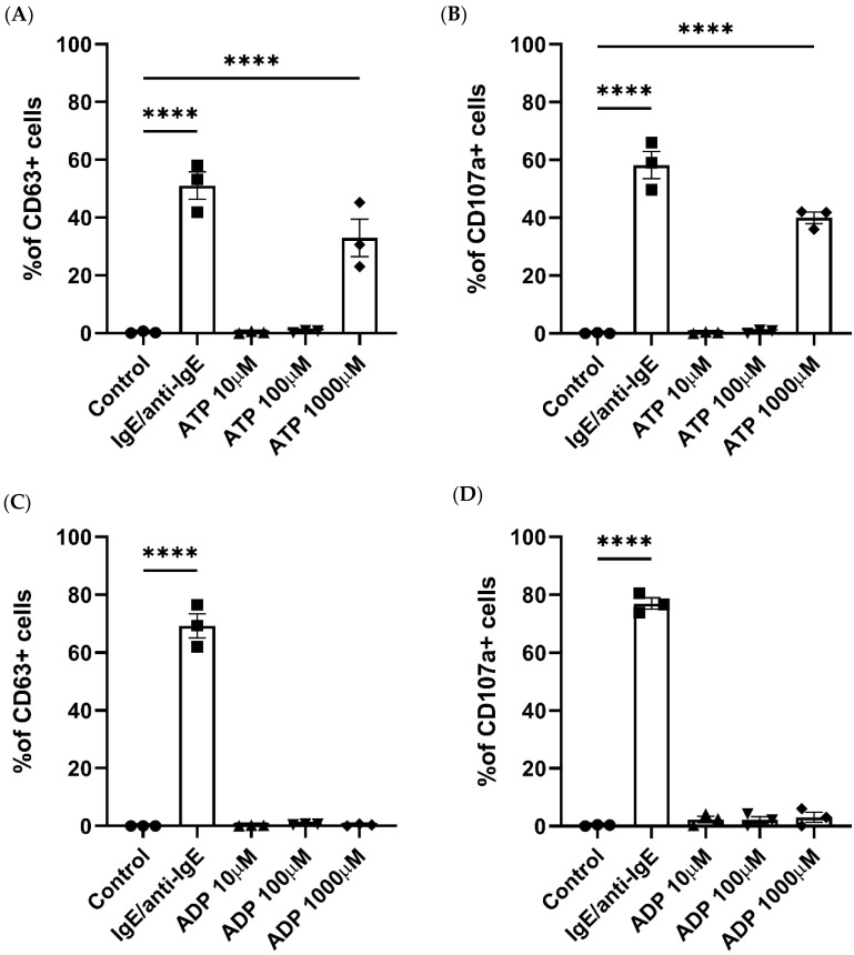
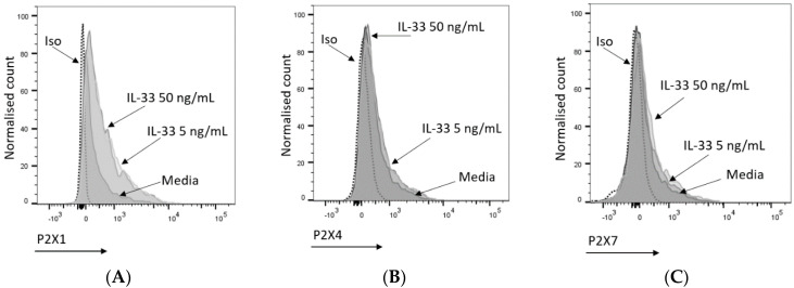
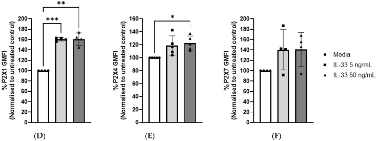
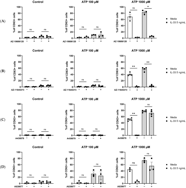
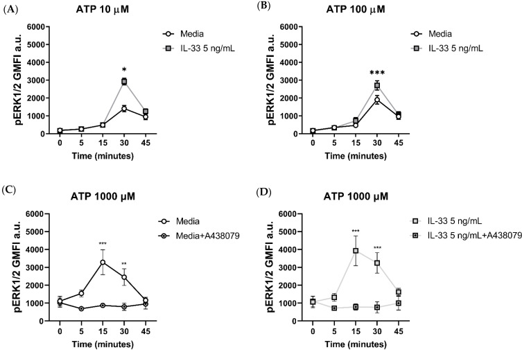
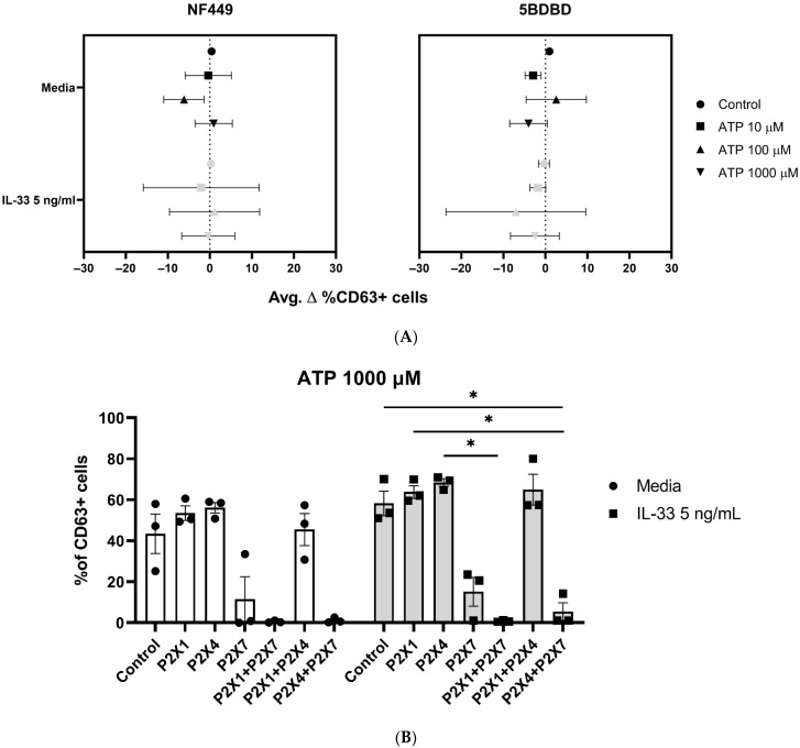
MCs are tissue-resident immune cells that strategically reside in barrier organs and respond effectively to a wide range of stimuli, such as IL-33, a mediator released upon epithelial damage. Adenosine triphosphate (ATP) accumulates at sites of tissue injury and is known to modulate MC activities. This study investigated how an inflammatory tissue environment rich in IL-33 modulates the ATP-mediated activation of MCs. Human primary MCs primed with IL-33 displayed a strongly increased response to ATP but not ADP. This resulted in increased degranulation, IL-8 release, and pERK1/2 signalling. Such effects are unique to IL-33 stimulation and not shared by the epithelial alarmin, TSLP. MC exposure to IL-33 also increased membrane expression of purinergic and ATP-binding P2X receptors. The use of selective P2X receptor inhibitors identified P2X7 receptor as the key mediator of the enhanced ATP-induced ERK1/2 signalling and degranulation in IL-33-primed MCs. Whilst the inhibition of P2X1 and P2X4 receptors had no effect on MC degranulation, inhibiting these receptors together with P2X7 resulted in further decreased MC-mediated degranulation. These data therefore point toward the potential mechanisms by which IL-33 contributes to the modulation of ATP-mediated activation in human MCs.

Keywords: ATP; IL-33; P2X1; P2X4; P2X7; mast cells.


Conflict of interest statement

The authors declare no conflicts of interest. The funders had no role in the design of the study; in the collection, analyses, or interpretation of data; in the writing of the manuscript; or in the decision to publish the results. K.A. is an employee of GSK; however, no conflicts of interest in relation to this manuscript have arisen.

Figures

Figure 1
Figure 1
Human mast cell degranulation is induced by ATP stimulation. Degranulation in response to ATP (A,B), ADP (C,D), IgE and anti-IgE (positive control), or a negative control was measured by the externalization of CD63 (A,C) or CD107a (B,D) and analysed using flow cytometry. Data are mean ± SEM of n = 3 experiments from individual MC cultures. Statistical differences are indicated; **** p < 0.0001 (ordinary one-way ANOVA with Šídák’s post hoc test).
Figure 2
Figure 2
IL-33 enhances ATP-mediated MC activities. MCs were pre-treated for 24 h with media control or IL-33 at the concentrations indicated, followed by activation with IgE/anti-IgE, ATP, or a negative control (media). (A) Degranulation was measured by cell staining with an anti-CD63 antibody (n = 3 separate experiments from separate MC cultures); (B) effect of treatment with IL-33 5 ng/mL (n = 3 experiments from separate MC cultures, left) and 50 ng/mL (n = 3 experiments from separate MC cultures, right) on ADP-induced MC degranulation measured by anti-CD63 antibody staining. (C) IL-8 cytokine secretion by IL-33-treated cells stimulated with ATP and ADP for 8 h (n = 6 independent experiments from six individual MC cultures). (D) Comparison of IL-33 and TSLP pre-treatments on ATP-induced cell degranulation as measured by CD63 flow cytometry staining (n = 4 separate experiments in separate MC cultures). Data are mean ± SEM. Statistical differences are indicated; ns: not significant, * p < 0.05, ** p < 0.01, *** p < 0.001, **** p < 0.0001 (two-way ANOVA with Šídák’s post hoc test).
Figure 3
Figure 3
Mast cell P2X receptor expression and modulation by IL-33. (AC) Representative histogram indicating P2X1 (A), P2X4 (B), and P2X7 (C) expression upon treatment with media (5 or 50 ng/mL IL-33 for 24 h). P2X1, P2X4, and P2X7 were stained and analysed by flow cytometry. The dotted lines indicate cell staining with isotype control antibodies. (DF) IL-33-treated or untreated MCs were stained for P2X1 (n = 2), P2X4 (n = 5), and P2X7 (n = 4); the geometric mean of fluorescence intensity (GMFI) was normalized to the negative control (untreated samples). Data are displayed as mean ± SEM. Statistical differences are indicated by * p < 0.05 and ** p < 0.01, *** p < 0.001 (one-way ANOVA with Šídák’s post hoc test).
Figure 3
Figure 3
Mast cell P2X receptor expression and modulation by IL-33. (AC) Representative histogram indicating P2X1 (A), P2X4 (B), and P2X7 (C) expression upon treatment with media (5 or 50 ng/mL IL-33 for 24 h). P2X1, P2X4, and P2X7 were stained and analysed by flow cytometry. The dotted lines indicate cell staining with isotype control antibodies. (DF) IL-33-treated or untreated MCs were stained for P2X1 (n = 2), P2X4 (n = 5), and P2X7 (n = 4); the geometric mean of fluorescence intensity (GMFI) was normalized to the negative control (untreated samples). Data are displayed as mean ± SEM. Statistical differences are indicated by * p < 0.05 and ** p < 0.01, *** p < 0.001 (one-way ANOVA with Šídák’s post hoc test).
Figure 4
Figure 4
Inhibition of P2X7 receptor by orthosteric and allosteric ligands in IL-33-treated mast cells. MCs were left untreated or incubated with IL-33 for 24 h at 5 ng/mL. The MCs were then exposed to 5 μM concentration of the allosteric P2X7 inhibitors AZ-10606120 ((A), n = 4) and AZ-11645373 ((B), n = 4) and 5 μM concentration of the orthosteric P2X7 inhibitors A438079 ((C), n = 3) and A804598 ((D), n = 3) for 15 min before subsequent ATP stimulation. Data are mean ± SEM. Statistical differences are indicated as follows: ns = no significance; * p < 0.05; ** p < 0.01 (one-way ANOVA with Šídák’s post hoc test).
Figure 5
Figure 5
Effect of the orthosteric P2X7 inhibitor on ERK 1/2 phosphorylation in IL-33-treated MCs stimulated with ATP. MCs were left untreated or incubated with IL-33 for 24 h at the concentrations indicated and stimulated with ATP concentrations of 10 µM (A) and 100 µM (B). ERK1/2 phosphorylation was measured flow cytometry using the geometric mean of fluorescence intensity (GMFI, n = 3 separate experiments in separate MC cultures). (C,D) The A438079 P2X7 inhibitor was added at a concentration of 5 μM to untreated (C) or IL-33-treated MCs (D) 15 min prior to stimulation with ATP 1000 µM (n = 4 separate experiments from individual MC cultures). Data are mean ± SEM. Statistical differences are indicated as follows: * p < 0.05, ** p < 0.01, *** p < 0.001 (two-way ANOVA with Sidak’s post hoc test).
Figure 6
Figure 6
The use of P2X1 and P2X4 receptors, alone or in combination, does not affect MC degranulation. MCs were left untreated or incubated with 5 ng/mL IL-33 for 24 h. (A) The MCs were exposed to NF449 (P2X1 inhibitor) and 5BDBD (P2X4 inhibitor) for 15 min before ATP stimulation at the concentrations indicated (n = 3 separate experiments). A statistical analysis showed no significance between the control, stimulated, and IL-33-primed cells. (B) The MCs were exposed to NF449 (P2X1 inhibitor), 5BDBD (P2X4 inhibitor), and A438079 (P2X7 inhibitor) alone or in combination for 15 min before 1000 µM ATP stimulation at the concentrations indicated (n = 3 separate experiments using different MC cultures). Statistical analysis showed no significant difference between the untreated controls and IL-33-primed cells. Data are presented as mean ± SEM. Statistical differences are indicated; * p < 0.05 (one-way ANOVA with Sidak’s post hoc test).

Similar articles

Cited by

References

    1. da Silva E.Z., Jamur M.C., Oliver C. Mast cell function: A new vision of an old cell. J. Histochem. Cytochem. 2014;62:698–738. doi: 10.1369/0022155414545334. - DOI - PMC - PubMed
    1. Lunderius-Andersson C., Enoksson M., Nilsson G. Mast Cells Respond to Cell Injury through the Recognition of IL-33. Front. Immunol. 2012;3:82. doi: 10.3389/fimmu.2012.00082. - DOI - PMC - PubMed
    1. Krystel-Whittemore M., Dileepan K.N., Wood J.G. Mast Cell: A Multi-Functional Master Cell. Front. Immunol. 2015;6:620. doi: 10.3389/fimmu.2015.00620. - DOI - PMC - PubMed
    1. Yu Y., Blokhuis B.R., Garssen J., Redegeld F.A. Non-IgE mediated mast cell activation. Eur. J. Pharmacol. 2016;778:33–43. doi: 10.1016/j.ejphar.2015.07.017. - DOI - PubMed
    1. Hara K.Y., Kondo A. ATP regulation in bioproduction. Microb. Cell Fact. 2015;14:198. doi: 10.1186/s12934-015-0390-6. - DOI - PMC - PubMed

MeSH terms

LinkOut - more resources