Review
. 2024 Feb 1;25(3):1734.
doi: 10.3390/ijms25031734.

In-Depth Genomic Analysis: The New Challenge in Congenital Heart Disease

Affiliations
Review

In-Depth Genomic Analysis: The New Challenge in Congenital Heart Disease

Francesco Nappi. Int J Mol Sci. .

Abstract

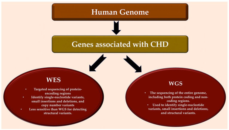
The use of next-generation sequencing has provided new insights into the causes and mechanisms of congenital heart disease (CHD). Examinations of the whole exome sequence have detected detrimental gene variations modifying single or contiguous nucleotides, which are characterised as pathogenic based on statistical assessments of families and correlations with congenital heart disease, elevated expression during heart development, and reductions in harmful protein-coding mutations in the general population. Patients with CHD and extracardiac abnormalities are enriched for gene classes meeting these criteria, supporting a common set of pathways in the organogenesis of CHDs. Single-cell transcriptomics data have revealed the expression of genes associated with CHD in specific cell types, and emerging evidence suggests that genetic mutations disrupt multicellular genes essential for cardiogenesis. Metrics and units are being tracked in whole-genome sequencing studies.

Keywords: congenital heart disease; copy number variants; de novo mutations; deletion; first heart field; gene variants; loss-of-function variant; second heart field; whole-exome sequencing; whole-genome sequencing.


Conflict of interest statement

The author declares no conflicts of interest.

Figures

Figure 1
Figure 1
The most frequent forms of CHD from the Paediatric Cardiac Genomics Consortium, with percentages indicating the prevalence of the malformation in the patient population. It is important to note that the percentages exceed 100% as a result of concomitant structural cardiac abnormalities in the individuals Abbreviations: AVCD, atrioventricular canal defect; CHD, congenital heart disease; CTM, conotruncal malformation; LVOTO, left ventricular outflow tract obstruction; SD, septal defect. Refs. [17,18].
Figure 2
Figure 2
A simplified scheme of CHD defects divided according to anatomical subtype and their corresponding oxygen saturation levels. Anomalies in the communication between the left and right ventricles, such as ASD or VSD or atrioventricular canal defects, usually result in acyanotic states due to a left-to-right shunting of oxygen-rich blood into the pulmonary circulation. LVOTOs can range from isolated aortic stenosis to a combination of anomalies, such as hypoplastic left heart syndrome with mitral and aortic stenosis, which can cause cyanosis. Conotruncal malformations, such as TOF and TGV, are often associated with cyanosis due to the shunting of deoxygenated blood into the systemic circulation. This information is from the Paediatric Cardiac Genomics Consortium. Abbreviations: Ao, aorta; LA, left atrium; LV, left ventricle; PA, pulmonary artery; RA, right atrium; RV, right ventricle; TGV, transposition of great vessel; TOF, tetralogy of Fallot. Refs. [17,18].
Figure 3
Figure 3
Detection of genes associated with CHD in human genome. Bioinformatic analytical tools such as WES and WGS platforms can be used to identify deleterious missense and LOF variants, small insertions or deletions, CNVs, and structural variants in large, aggregated, population-based sequencing datasets. Abbreviations: CHD, congenital heart disease; CNV, copy number variant; LOF, loss-of-function; WES, whole-exome sequencing; WGS, whole-genome sequencing. Refs. [18,19,20,21,22,23,24,25].
Figure 4
Figure 4
This pie chart illustrates the various causes of CHD and their relative proportions in contributing to the disease. Zaidi et al. [36] were the first to estimate the contribution of de novo mutations to CHD, while Jin et al. [22] presented the first model to systematically investigate inherited CHD mutations. Recessive transmitted causes involve variants in cilia and known CHD genes. Dominant causes of TOF include LOFs associated with FLT4, KDR, TBX1, and NOTCH1. DNMs may involve chromatin modifiers (2%) and dominant CHD genes (6%). Aneuploidy aetiology is related to trisomy 13, 18, or 21 and monosomy X. Non-genetic causes of CHD include teratogenic exposures such as alcohol; antiseizure and antiretroviral medication; and illnesses and infectious agents, such as diabetes, hypercholesterolemia, and rubella. The causes of CHD are investigated through gene–gene interactions, gene–environment interactions, polygenic inheritance, and epigenetics. CNVs, including Del22q11.2, Del20p12, Del17q11, Del11q24-25, Del8p23.1, Del1q21.1, Dup12q24, and Dup7q11.23, are also considered. Non-genetic causes of CHD are discussed in References [23,37,38,39,40]. Abbreviations: CHD, congenital heart disease; CNVs, copy number variations; Del, deletion, DMSs, de novo mutations; Dup, duplication; LOF, loss of function.
Figure 5
Figure 5
This schematic illustrates the embryonic development of the human heart, including first and second heart field formation, heart tube formation and pumping, looping, neural crest migration, and septation. The process results in a fully developed heart at the end of gestation. Abbreviation: HF, heart field. Refs. [41,42,43,44,45].
Figure 6
Figure 6
The left diagram shows that two types of congenital heart defects (ASD and VSD) are linked to gene mutations in transcription factors and signalling molecules. The right diagram illustrates abnormal connections between the left and right chambers of the heart, including atrial or ventricular septal defects or atrioventricular canal defects. These defects typically do not result in cyanosis because oxygenated blood moves from the left to the right side of the heart and into the pulmonary circulation. Abbreviations: ASD, atrial septal defect; VSD, ventricular septal defect. The image was created using modified images licensed under a Creative Commons Attribution 3.0 Unported License. Refs. [88,89].
Figure 7
Figure 7
Fused bicuspid aortic valve. Panel (A) represents a short-axis normal tricuspid aortic pattern with anatomical proximities. Panels (B,C) represent two cusp fusion patterns seen in the short heart axis. All BAVs have three sinuses. Raphe structure is between the fused cusps. Non-fused cusp is prominent in respect to the fused ones. (B) Left-noncoronary fusion pattern; (C) right-noncoronary fusion pattern. The commissure angle of the non-fused cusp has a degree < 180°. Abbreviations: L, left coronary sinus; LA, left atrium; LC, left cusp; LCA, left coronary artery; MV, mitral valve; N, noncoronary sinus; NC, noncoronary cusp; PV, pulmonary valve; R, right coronary sinus; RC, right cusp; RCA, right coronary artery; RV, right ventricle, TV, tricuspid valve. Licenses Centre Cardiologique du Nord (with permission). License Number 5644110549132 License date 8 October 2023; publication NEJM; Title: Mitral valve Repair for Mitral valve prolapse. Ref [91].
Figure 8
Figure 8
Two examples of left-sided obstructive lesions, represented by a normal and a pathological aortic valve with the corresponding surgical treatment and an aortic coarctation with the corresponding surgical approach. Top left: Foetal aortic valve stenosis is a heart condition where the aortic valve, located between the left ventricle and aorta, cannot fully open. This causes blood to flow out of the left ventricle and enlarges the right ventricle, which can lead to poor foetal growth due to changes in blood flow. Bottom left: The procedure performed is an extended 3-patch supravalvular aortic stenosis repair. (AC) In this procedure, the ascending aorta is transected at its narrowest point, and three incisions are made into the sinuses of Valsalva. (B) The sinuses are then enlarged using three pericardial patches, and the patch from the noncoronary sinus is extended into the ascending aorta to ensure symmetrical enlargement of the narrow segment. (C) Top and bottom right: The diagram illustrates the surgical procedure employed to treat severe aortic coarctation. Extended resection is performed, followed by end-to-end anastomosis. Abbreviations: PDA, patent ductus arteriosus; L.C.C, left common carotid artery; L.S.A, left subclavian artery. The image was created using modified images licensed under a Creative Commons Attribution 3.0 Unported License. Refs. [89,92].
Figure 9
Figure 9
The image shows a comparison between a normal heart (bottom) and a heart (top) affected by hypoplastic left heart syndrome. The different treatment strategies are represented. On the left: Diagram of hypoplastic left heart syndrome. The mitral and aortic valves, left ventricular cavity, and aorta are severely underdeveloped, resulting in systemic blood flow being supplied by the patent ductus arteriosus. On the right: Staged reconstruction for hypoplastic left heart syndrome. (AF) Stage I of the Norwood reconstruction involves using a modified Blalock–Taussig (BT) shunt. (A) The second stage is the Norwood reconstruction or the Norwood reconstruction using the Sano modification. (B) A hybrid procedure is also an option. (C,D). The final stage is the Fontan procedure. (E,F) LA stands for left atrium. LPA represents left pulmonary artery. LV stands for left ventricle. RA denotes right atrium. RPA represents right pulmonary artery. RV stands for right ventricle. SVC denotes superior vena cava. The image was created using modified images licensed under a Creative Commons Attribution 3.0 Unported License. Ref. [89].
Figure 10
Figure 10
(A) A Venn diagram that shows the overlap of genes associated with LSOLs based on disease subtype. Genes without robust evidence of association are marked with an asterisk. (B) The genetic landscape of left-sided obstructive lesions. The genes marked with an asterisk have no robust evidence of association. The genes code for transcription factors, structural or contractile proteins, and cell signalling components. Abbreviations: AS, aortic stenosis; BAV, bicuspid aortic valve; CoA, coarctation of the aorta; HLHS, hyperplastic left heart syndrome; IAA, interrupted aortic arch; LSOL, left-sided obstructive lesion. The image was created using modified images licensed under a Creative Commons Attribution 3.0 Unported License. Asterisk denotes genes lacking robust evidence of association. Genes code for transcription factors, structural or contractile proteins, § cell signaling components.
Figure 11
Figure 11
Germline mutations that activate RAS/MAPK signalling cause “RASopathies”. These are a group of rare human developmental disorders affecting over 400,000 individuals in the United States alone. Costello syndrome (CS) is a syndrome of multiple congenital anomalies. It is caused by heterozygous activating germline mutations in HRAS [1]. HRAS hyperactivation inhibits the LKB1/AMPK pathway. Most individuals with CS have a mutation in HRAS at position G12, with over 80% having a p.G12S substitution. Children with CS typically present with increased birth weight, dysmorphic craniofacial features, failure to thrive, and gastroesophageal reflux with oral aversion, especially in the neonatal period. CS can also affect the skin, causing excessive wrinkling and redundancy over the dorsum of the hands and feet, and deep plantar and palmar folds. In addition, individuals with CS have an increased risk of developing benign or malignant tumours. miR-221*, miR-221-5p; +++, increased HRAS-GTP function Ref [115].
Figure 12
Figure 12
The diagram illustrates a type of obstructive lesion on the right side. (A) PA/IVS is an abnormality in which there is complete closure of the communication from the RV to the PA and no ventricular septal defect. (B) PA/IVS is a disorder with varying morphology. The RV is often small or hypoplastic, or it can be significantly dilated, as seen in association with Ebstein’s anomaly or TV dysplasia and severe TV regurgitation. Although extracardiac anomalies are unusual in PA/IVS, many different cardiac anomalies can be observed. PA/IVS is typically associated with abnormalities of the right side of the heart, including dysplastic TV leaflets and thickened chordae, which may cause severe regurgitation in utero. Atrial septal defects are also common. It is worth noting that the TV annulus is often small in many cases. In some cases, the tricuspid valve may be fully sealed off, causing tricuspid atresia, or, more commonly, it may be moderately to severely underdeveloped, resulting in tricuspid stenosis. Especially in the setting of a hypertrophied, hypertensive RV with little or no tricuspid regurgitation, coronary abnormalities are common in PA/IVS. An inverse relationship exists between RV size and coronary anomalies—the smaller the RV and tricuspid annulus, the greater the likelihood of coronary anomalies. Abbreviations: PAPA/IVS, pulmonary atresia with intact ventricular septum; PA, pulmonary artery; RV, right ventricle; TV, tricuspid valve. The image was created using modified images licensed under a Creative Commons Attribution 3.0 Unported License. Ref. [89].
Figure 13
Figure 13
Conotruncal defects include conditions such as tetralogy of Fallot, persistent truncus arteriosus, and transposition of the great arteries, which are caused by the underdevelopment or malposition of the ventricular septum, outflow tract, and/or great arteries. Conotruncal malformations often result in cyanosis due to the shunting of deoxygenated blood into the systemic circulation. The image was created using modified images licensed under a Creative Commons Attribution 3.0 Unported License. Ref. [89].

Similar articles

Cited by

References

    1. Ibrahim S., Gaborit B., Lenoir M., Collod-Beroud G., Stefanovic S. Maternal Pre-Existing Diabetes: A Non-Inherited Risk Factor for Congenital Cardiopathies. Int. J. Mol. Sci. 2023;24:16258. doi: 10.3390/ijms242216258. - DOI - PMC - PubMed
    1. Dilli D., Akduman H., Zenciroğlu A., Çetinkaya M., Okur N., Turan Ö., Özlü F., Çalkavur Ş., Demirel G., Koksal N., et al. Neonatal Outcomes of Critical Congenital Heart Defects: A Multicenter Epidemiological Study of Turkish Neonatal Society: Neonatal Outcomes of CCHD. Pediatr. Cardiol. 2024;45:257–271. doi: 10.1007/s00246-023-03362-z. - DOI - PubMed
    1. Hossin M.Z., de la Cruz L.F., McKay K.A., Oberlander T.F., Sandström A., Razaz N. Association of pre-existing maternal cardiovascular diseases with neurodevelopmental disorders in offspring: A cohort study in Sweden and British Columbia, Canada. Int. J. Epidemiol. 2023:dyad184. doi: 10.1093/ije/dyad184. - DOI - PMC - PubMed
    1. Bakker M.K., Bergman J.E.H., Krikov S., Amar E., Cocchi G., Cragan J., de Walle H.E.K., Gatt M., Groisman B., Liu S., et al. Prenatal diagnosis and prevalence of critical congenital heart defects: An international retrospective cohort study. BMJ Open. 2019;9:e028139. doi: 10.1136/bmjopen-2018-028139. - DOI - PMC - PubMed
    1. Hoffman J.I.E., Kaplan S. The incidence of congenital heart disease. J. Am. Coll. Cardiol. 2002;39:1890–1900. doi: 10.1016/S0735-1097(02)01886-7. - DOI - PubMed