. 2024 Feb 1;25(3):1789.
doi: 10.3390/ijms25031789.

Clinical Characteristics and Local Histopathological Modulators of Endometriosis and Its Progression

Affiliations

Clinical Characteristics and Local Histopathological Modulators of Endometriosis and Its Progression

Anca-Maria Istrate-Ofiţeru et al. Int J Mol Sci. .

Abstract

Endometriosis (E) and adenomyosis (A) are associated with a wide spectrum of symptoms and may present various histopathological transformations, such as the presence of hyperplasia, atypia, and malignant transformation occurring under the influence of local inflammatory, vascular and hormonal factors and by the alteration of tumor suppressor proteins and the inhibition of cell apoptosis, with an increased degree of lesion proliferation.

Material and methods: This retrospective study included 243 patients from whom tissue with E/A or normal control uterine tissue was harvested and stained by histochemical and classical immunohistochemical staining. We assessed the symptomatology of the patients, the structure of the ectopic epithelium and the presence of neovascularization, hormone receptors, inflammatory cells and oncoproteins involved in lesion development. Atypical areas were analyzed using multiple immunolabeling techniques.

Results: The cytokeratin (CK) CK7+/CK20- expression profile was present in E foci and differentiated them from digestive metastases. The neovascularization marker cluster of differentiation (CD) 34+ was increased, especially in areas with malignant transformation of E or A foci. T:CD3+ lymphocytes, B:CD20+ lymphocytes, CD68+ macrophages and tryptase+ mast cells were abundant, especially in cases associated with malignant transformation, being markers of the proinflammatory microenvironment. In addition, we found a significantly increased cell division index (Ki67+), with transformation and inactivation of tumor suppressor genes p53, B-cell lymphoma 2 (BCL-2) and Phosphatase and tensin homolog (PTEN) in areas with E/A-transformed malignancy.

Conclusions: Proinflammatory/vascular/hormonal changes trigger E/A progression and the onset of cellular atypia and malignant transformation, exacerbating symptoms, especially local pain and vaginal bleeding. These triggers may represent future therapeutic targets.

Keywords: adenomyosis; endometriosis; multiple immunohistochemistry.


Conflict of interest statement

The authors declare no conflicts of interest.

Figures
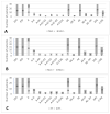
Chart 1
Chart 1
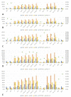
(AE) Number of patients with Met (1A)/Men (1B)/PI (1C)/SI (1D)/BD (1E); EPP: endometrium proliferative phase; ESP: endometrium secretary phase; A: adenomyosis; A-H: adenomyosis with hyperplasia without atypia; A-AH: adenomyosis with hyperplasia with atypia; A-EC-G1: adenomyosis associated with endometrioid carcinoma G1; A-EC-G2: adenomyosis associated with endometrioid carcinoma G2; A-EC-G3: adenomyosis associated with endometrioid carcinoma G3; A-CCAC: adenomyosis associated with clear cell adenocarcinoma; OE: ovarian endometriosis; OE-A: ovarian endometriosis with cell atypia; AE: abdominal endometriosis; AE-H: abdominal hyperplastic endometriosis without atypia; AE-AH: abdominal hyperplastic endometriosis with atypia; DIE: deep infiltrative endometriosis; C-DIE: colorectal DIE; Met: metrorrhagia; Men: menorrhagia; PI: primary infertility; SI: secondary infertility; BD: bowel disorder.
Chart 1
Chart 1
(AE) Number of patients with Met (1A)/Men (1B)/PI (1C)/SI (1D)/BD (1E); EPP: endometrium proliferative phase; ESP: endometrium secretary phase; A: adenomyosis; A-H: adenomyosis with hyperplasia without atypia; A-AH: adenomyosis with hyperplasia with atypia; A-EC-G1: adenomyosis associated with endometrioid carcinoma G1; A-EC-G2: adenomyosis associated with endometrioid carcinoma G2; A-EC-G3: adenomyosis associated with endometrioid carcinoma G3; A-CCAC: adenomyosis associated with clear cell adenocarcinoma; OE: ovarian endometriosis; OE-A: ovarian endometriosis with cell atypia; AE: abdominal endometriosis; AE-H: abdominal hyperplastic endometriosis without atypia; AE-AH: abdominal hyperplastic endometriosis with atypia; DIE: deep infiltrative endometriosis; C-DIE: colorectal DIE; Met: metrorrhagia; Men: menorrhagia; PI: primary infertility; SI: secondary infertility; BD: bowel disorder.
Figure 1
Figure 1
(AC): Morphological aspects of E/A lesions in classical HE staining. (A): Endometrial glands (white arrowhead) and peri-glandular stroma (black arrow) are visualized in the myometrial structure; (B): DIE lesions—ectopic presence of endometrial glands (white arrowhead) in the peritoneal structure with stromal and inflammatory infiltrate (black arrow) in the sub-mesothelial connective tissue; (C): C-DIE lesions, endometrial glands (white arrowhead) are seen in the colonic submucosa structure, [HE staining, ×100]. HE: hematoxylin–eosin; A: adenomyosis; DIE: deep infiltrative endometriosis; C-DIE: colorectal deep infiltrative endometriosis.
Figure 2
Figure 2
(A,B): Labeling of cytokeratins in E/A lesions (A): A foci (white arrowhead) are observed in the myometrial structure. Immunostaining with anti-CK7 antibody (brown label), [×200]; (B): Diffuse immunostaining of the glandular epithelium with anti-CK7 antibody is observed in the A-AH foci (white arrowhead), [×200]. A: adenomyosis; A-AH: uterine endometriosis with hyperplasia with atypia; CK7: cytokeratin 7.
Figure 3
Figure 3
(A,B): Labeling of hormone receptors in A/E. (A): Immunostaining with anti-ERα antibody in A (brown marking, white arrowhead), [×200]; (B): the presence of endometrial epithelium, peri-glandular stroma and PR (brown marking) is observed in the structure of the colonic mucosa. Immunostaining with anti-PRα antibody (white arrowhead), [×200]. ERα: estrogen receptor alpha; PRα: progesterone receptor alpha; A: adenomyosis; C-DIE: colorectal deep infiltrative endometriosis.
Figure 4
Figure 4
Labeling of stroma (white arrowhead) in A-H. Immunostaining with anti-CD10 antibody, [×100]. A-H: adenomyosis with hyperplasia without atypia; CD10: cluster of differentiation 10.
Figure 5
Figure 5
(A,B): Vascularization in ectopic foci of E/A. (A): AE lesions stained with anti-CD34 antibody highlighting the capillary endothelium of newly formed perilesional blood vessels (white arrowhead), [×400]; (B): A-AH lesion immunolabeled with anti-CD34 antibody highlighting the capillary endothelium of newly formed perilesional blood vessels (white arrowhead), [×400].
Chart 2
Chart 2
Distribution of mean values of CD34+ neoformation vessels and inflammatory system cells highlighting CD3+ T lymphocytes, CD20+ B lymphocytes, CD68+ macrophages and tryptase+ mast cells. The number of vessels exhibited overall differences between the studied groups regarding CD34: [F(15,242) = 450.601, p < 0.05]; CD3: [F(15,242) = 1970.346, p < 0.05]; CD20: [F(15,242) = 459.200, p < 0.05]; CD68: [F(15,242) = 417.152, p < 0.05]; tryptase: [F(15,242) = 457.346, p < 0.05]. CD: cluster of differentiation; EPP: endometrium proliferative phase; ESP: endometrium secretory phase; A: adenomyosis; A-H: adenomyosis with hyperplasia without atypia; A-AH: adenomyosis with hyperplasia with atypia; A-EC-G1: adenomyosis associated with endometrioid carcinoma G1; A-EC-G2: adenomyosis associated with endometrioid carcinoma G2; A-EC-G3: adenomyosis associated with endometrioid carcinoma G3; A-CCAC: adenomyosis associated with clear cell adenocarcinoma; OE: ovarian endometriosis; OE-A: ovarian endometriosis with cell atypia; AE: abdominal wall endometriosis; AE-H: abdominal wall hyperplastic endometriosis without atypia; AE-AH: abdominal wall hyperplastic endometriosis with atypia; DIE: deep infiltrative endometriosis; C-DIE; colorectal deep infiltrative endometriosis.
Figure 6
Figure 6
(A,B): Immunohistochemical aspects of E/A lesions stained with anti-CD3 antibody. (A): AE lesion immunolabeled with anti-CD3 antibody highlighting T lymphocytes (white arrowhead), [×400]. (B): Focus of A-EC-G2 immunolabeled with anti-CD3 antibody highlighting T lymphocytes (white arrowhead), [×400]. AE: abdominal wall endometriosis; A-EC-G2: uterine endometriosis associated with endometrioid carcinoma G2.
Figure 7
Figure 7
(A,B): Immunohistochemical aspects of E/A lesions stained with anti-CD20 antibody. M: OE lesion immunolabeled with anti-CD20 antibody highlighting B lymphocytes (white arrowhead), [×400]; N: A-AH lesion immunolabeled with anti-CD20 antibody highlighting B lymphocytes (white arrowhead), [×400]. OE: ovarian endometriosis; A-AH: uterine endometriosis with hyperplasia with atypia.
Figure 8
Figure 8
Immunohistochemical aspects of E lesions in staining with anti-CD68 antibody. O: OE lesion immunolabeled with anti-CD68 antibody highlighting macrophages (white arrowhead), [×400]. OE: ovarian endometriosis; CD68: cluster of differentiation 68.
Figure 9
Figure 9
(A,B): Immunohistochemical aspects of E lesions stained with anti-tryptase antibody. (A): C-DIE lesion immunolabeled with anti-tryptase antibody highlighting mast cells (white arrowhead), [×400]. (B): A-AH colorectal DIE lesion immunolabeled with anti-tryptase antibody highlighting mast cells (white arrowhead), [×400]. C-DIE: colorectal deep infiltrative endometriosis; A-AH: uterine endometriosis with hyperplasia with atypia.
Figure 10
Figure 10
(A,B): Immunohistochemical aspects of E/A lesions stained with anti-Ki67/p53 antibody. (A): A-EC-G2 lesion immunolabeled with anti-Ki67 antibody highlighting dividing cells (white arrowhead), [×400]. (B): A-AH lesion immunolabeled with anti-p53 antibody (white arrowhead), [×400]. A-EC-G2: uterine endometriosis associated with endometrioid carcinoma G2; Ki67: marker of proliferation Ki 67; p53: tumor protein 53.
Figure 11
Figure 11
(A,B): Immunohistochemical aspects of E/A lesions stained with anti-BCL-/PTEN antibody. (A): A-AH lesion immunolabeled with anti-BCL-2 antibody (white arrowhead), [×400]. (B): A-EC-G2 lesion immunolabeled with anti-PTEN antibody (white arrowhead), [×400]. A-AH: uterine endometriosis with hyperplasia with atypia; A-EC-G2: uterine endometriosis associated with endometrioid carcinoma G2; BCL-2: B cell lymphoma 2; PTEN: phosphatase and tensin homolog.
Chart 3
Chart 3
(AE): Associations of growth tendencies between inflammatory cells, neovascularization and symptomatology: Met (A), Men (B), PI (C), SI (D), BD (E). EPP: endometrium proliferative phase; ESP: endometrium secretary phase; A: adenomyosis; A-H: adenomyosis with hyperplasia without atypia; A-AH: adenomyosis with hyperplasia with atypia; A-EC-G1: adenomyosis associated with endometrioid carcinoma G1; A-EC-G2: adenomyosis associated with endometrioid carcinoma G2; A-EC-G3: adenomyosis associated with endometrioid carcinoma G3; A-CCAC: adenomyosis associated with clear cell adenocarcinoma; OE: ovarian endometriosis; OE-A: ovarian endometriosis with cell atypia; AE: abdominal wall endometriosis; AE-H: abdominal wall hyperplastic endometriosis without atypia; AE-AH: abdominal wall hyperplastic endometriosis with atypia; DIE: deep infiltrative endometriosis; C-DIE; colorectal deep infiltrative endometriosis; CD: cluster of differentiation; Met: metrorrhagia; Men: menorrhagia; PI: primary infertility; SI: secondary infertility; BD: bowel disorders.
Chart 4
Chart 4
(AE) Trends between pain intensity and pathology location. (A): VAS 0->1; (B): VAS 2->3; (C): VAS 4->5; (D): VAS 6->7; (E): VAS 8->10. EPP: endometrium proliferative phase; ESP: endometrium secretary phase; A: adenomyosis; A-H: adenomyosis with hyperplasia without atypia; A-AH: adenomyosis with hyperplasia with atypia; A-EC-G1: adenomyosis associated with endometrioid carcinoma G1; A-EC-G2: adenomyosis associated with endometrioid carcinoma G2; A-EC-G3: adenomyosis associated with endometrioid carcinoma G3; A-CCAC: adenomyosis associated with clear cell adenocarcinoma; OE: ovarian endometriosis; OE-A: ovarian endometriosis with cell atypia; AE: abdominal wall endometriosis; AE-H: abdominal wall hyperplastic endometriosis without atypia; AE-AH: abdominal wall hyperplastic endometriosis with atypia; DIE: deep infiltrative endometriosis; C-DIE; colorectal deep infiltrative endometriosis; CD: cluster of differentiation; VAS: visual analogue scale.

References

    1. Schorage J.O., Schaffer J.J., Halvorson L.M., Hoffman B.L., Bradshaw K.D., Cunningham F.G. Williams Gynecology. McGraw Hill Medical; New York, NY, USA: 2008.
    1. Zhai J., Vannuccini S., Petraglia F., Giudice L.C. Adenomyosis: Mechanisms and Pathogenesis. Semin. Reprod. Med. 2020;38:129–143. doi: 10.1055/s-0040-1716687. - DOI - PMC - PubMed
    1. Ibrahim M.G., Chiantera V., Frangini S., Younes S., Köhler C., Taube E.T., Plendl J., Mechsner S. Ultramicro-Trauma in the Endometrial-Myometrial Junctional Zone and Pale Cell Migration in Adenomyosis. Fertil. Steril. 2015;104:1475–1483.e1-3. doi: 10.1016/j.fertnstert.2015.09.002. - DOI - PubMed
    1. Leyendecker G., Herbertz M., Kunz G., Mall G. Endometriosis Results from the Dislocation of Basal Endometrium. Hum. Reprod. 2002;17:2725–2736. doi: 10.1093/humrep/17.10.2725. - DOI - PubMed
    1. Struble J., Reid S., Bedaiwy M.A. Adenomyosis: A Clinical Review of a Challenging Gynecologic Condition. J. Minim. Invasive Gynecol. 2016;23:164–185. doi: 10.1016/j.jmig.2015.09.018. - DOI - PubMed

Substances