. 2024 Feb 2;25(3):1816.
doi: 10.3390/ijms25031816.

Investigating the Role of 17-Beta Estradiol in the Regulation of the Unfolded Protein Response (UPR) in Pancreatic Beta Cells

Affiliations

Investigating the Role of 17-Beta Estradiol in the Regulation of the Unfolded Protein Response (UPR) in Pancreatic Beta Cells

Monica De Paoli et al. Int J Mol Sci. .

Abstract

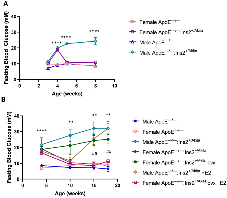
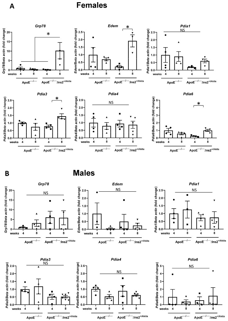
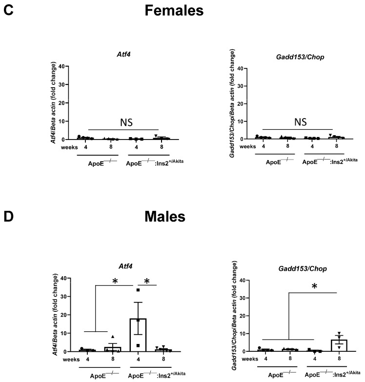
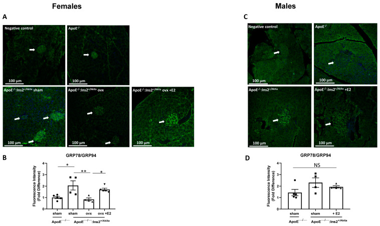
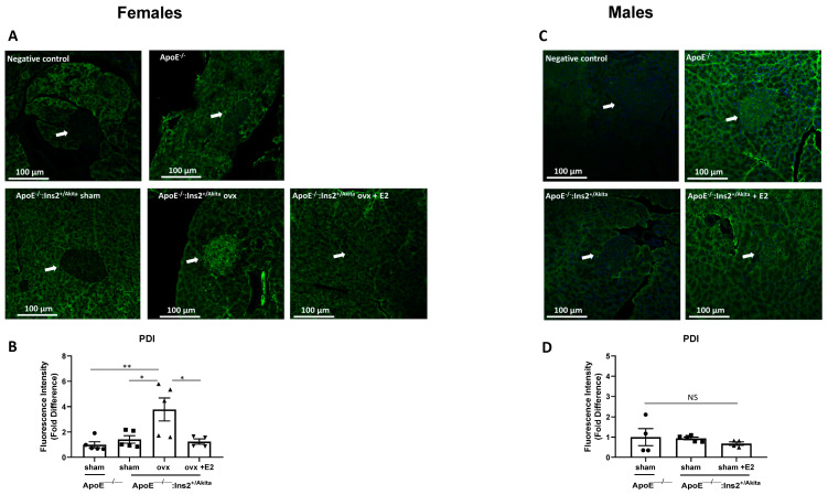
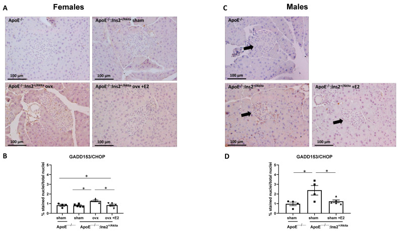
Diabetes mellitus is clinically defined by chronic hyperglycemia. Sex differences in the presentation and outcome of diabetes exist with premenopausal women having a reduced risk of developing diabetes, relative to men, or women after menopause. Accumulating evidence shows a protective role of estrogens, specifically 17-beta estradiol, in the maintenance of pancreatic beta cell health; however, the mechanisms underlying this protection are still unknown. To elucidate these potential mechanisms, we used a pancreatic beta cell line (BTC6) and a mouse model of hyperglycemia-induced atherosclerosis, the ApoE-/-:Ins2+/Akita mouse, exhibiting sexual dimorphism in glucose regulation. In this study we hypothesize that 17-beta estradiol protects pancreatic beta cells by modulating the unfolded protein response (UPR) in response to endoplasmic reticulum (ER) stress. We observed that ovariectomized female and male ApoE-/-:Ins2+/Akita mice show significantly increased expression of apoptotic UPR markers. Sham operated female and ovariectomized female ApoE-/-:Ins2+/Akita mice supplemented with exogenous 17-beta estradiol increased the expression of adaptive UPR markers compared to non-supplemented ovariectomized female ApoE-/-:Ins2+/Akita mice. These findings were consistent to what was observed in cultured BTC6 cells, suggesting that 17-beta estradiol may protect pancreatic beta cells by repressing the apoptotic UPR and enhancing the adaptive UPR activation in response to pancreatic ER stress.

Keywords: 17-beta estradiol; diabetes; endoplasmic reticulum stress; pancreatic beta cells; unfolded protein response.


Conflict of interest statement

The authors declare no conflicts of interest. The funders had no role in the design of the study; in the collection, analyses, or interpretation of data; in the writing of the manuscript; or in the decision to publish the results.

Figures

Figure 1
Figure 1
(A) Fasting blood glucose levels are lower in female ApoE−/−:Ins2+/Akita mice, compared to age-matched male ApoE−/−:Ins2+/Akita mice. Fasting blood glucose levels were determined in female and age-matched male ApoE−/− and ApoE−/−:Ins2+/Akita. n = 4–7 mice per experimental group; female male ApoE−/−:Ins2+/Akita mice. Bars represent standard error of the mean (SEM). (B) Ovariectomy increases and estrogen supplementation decreases fasting blood glucose levels in female ApoE−/−:Ins2+/Akita mice. Fasting blood glucose levels were determined in sham operated female, ovariectomized (ovx) female, ovx female supplemented with 17-beta estradiol (E2), age-matched male ApoE−/−:Ins2+/Akita mice supplemented with estrogen or not, and male and female ApoE−/− controls. A transient reduction in blood glucose was observed in age-matched male ApoE−/−:Ins2+/Akita mice supplemented with estrogen. n = 5 mice per experimental group. **** p < 0.0001 male ApoE−/−:Ins2+/Akita mice vs. age matched male and female ApoE−/− controls. ** p < 0.01 male ApoE−/−:Ins2+/Akita mice vs. age matched sham operated female ApoE−/−:Ins2+/Akita mice. ## p < 0.01 ovx female ApoE−/−:Ins2+/Akita mice vs. ovx female ApoE−/−:Ins2+/Akita mice + E2. Bars represent standard error of the mean (SEM).
Figure 2
Figure 2
Expression of adaptive and apoptotic UPR markers in RNA transcripts of isolated pancreatic islets. Transcripts from isolated pancreatic islets were analyzed for the expression of the adaptive UPR markers Grp78, Edem, Pdia1, Pdia3, Pdia4, Pdia6 in (A) female and (B) male ApoE−/− and ApoE−/−:Ins2+/Akita mice at 4 and 8 weeks of age. Expression of the apoptotic UPR markers Gadd153/Chop and Atf4 in transcripts from isolated pancreatic islets of female (C) and male (D) ApoE−/− and ApoE−/−:Ins2+/Akita mice at 4 and 8 weeks of age. RNA transcripts isolated from pancreata from female ApoE−/−:Ins2+/Akita mice show significantly increased expression of adaptive UPR markers, while transcripts from male ApoE−/−:Ins2+/Akita mice show a significant increase in apoptotic UPR markers. Each sample represents pooled islets from n = 4–8 mice. n = 3–6 samples per experimental group, analyzed in technical duplicates. * p < 0.05, NS, not significant. Bars represent standard error of the mean (SEM).
Figure 2
Figure 2
Expression of adaptive and apoptotic UPR markers in RNA transcripts of isolated pancreatic islets. Transcripts from isolated pancreatic islets were analyzed for the expression of the adaptive UPR markers Grp78, Edem, Pdia1, Pdia3, Pdia4, Pdia6 in (A) female and (B) male ApoE−/− and ApoE−/−:Ins2+/Akita mice at 4 and 8 weeks of age. Expression of the apoptotic UPR markers Gadd153/Chop and Atf4 in transcripts from isolated pancreatic islets of female (C) and male (D) ApoE−/− and ApoE−/−:Ins2+/Akita mice at 4 and 8 weeks of age. RNA transcripts isolated from pancreata from female ApoE−/−:Ins2+/Akita mice show significantly increased expression of adaptive UPR markers, while transcripts from male ApoE−/−:Ins2+/Akita mice show a significant increase in apoptotic UPR markers. Each sample represents pooled islets from n = 4–8 mice. n = 3–6 samples per experimental group, analyzed in technical duplicates. * p < 0.05, NS, not significant. Bars represent standard error of the mean (SEM).
Figure 3
Figure 3
Expression of adaptive UPR markers GRP78/94 in pancreatic sections. Immunofluorescence staining was performed to measure the expression of adaptive UPR markers GRP78/GRP94 in pancreatic islet sections of sham-operated, ovariectomized, ovariectomized supplemented with 17-beta estradiol (E2) female (A,B) ApoE−/−:Ins2+/Akita and female ApoE−/− mice; and male (C,D) male ApoE−/−:Ins2+/Akita supplemented with estrogen (E2) or not. Ovariectomy significantly reduces the expression of GRP78/94 in female ApoE−/−:Ins2+/Akita mice, while no significant differences are observed in male experimental groups. n = 4–5 per group. * p < 0.05, ** p < 0.01, NS, not significant. Bars represent standard error of the mean (SEM).
Figure 4
Figure 4
Expression of adaptive UPR marker PDI in pancreatic islets. Immunofluorescent staining was performed to evaluate the expression of adaptive UPR marker PDI in pancreatic islet sections of sham-operated, ovariectomized, ovariectomized supplemented with 17-beta estradiol (E2) female (A,B) ApoE−/−:Ins2+/Akita and age matched female ApoE−/− mice; and male (C,D) male ApoE−/−:Ins2+/Akita supplemented with 17-beta estradiol or not. n = 4–5 per group. * p < 0.05, ** p < 0.01, NS, not significant. Bars represent standard error of the mean (SEM).
Figure 5
Figure 5
Expression of apoptotic UPR marker GADD153/CHOP in pancreatic sections. Immunostaining was performed to measure the expression of apoptotic UPR marker GADD153/CHOP in pancreatic islet sections of sham-operated, ovariectomized, ovariectomized supplemented with 17-beta estradiol (E2) female (A,B) ApoE−/−:Ins2+/Akita and female ApoE−/− mice; and male (C,D) ApoE−/−:Ins2+/Akita supplemented with estrogen (E2) or not, compared to age matched ApoE−/− controls. Data are expressed as percentage of brown-stained nuclei versus total nuclei in the islet. Ovariectomy significantly increases the expression of the apoptotic marker GADD153/CHOP in female ApoE−/−:Ins2+/Akita mice to levels similar to those observed in male ApoE−/−:Ins2+/Akita mice. n = 4–5 per group. * p < 0.05, Bars represent standard error of the mean (SEM).
Figure 6
Figure 6
Expression of Grp78 and Gadd153/Chop in BTC6 cells pretreated with estrogen. Transcripts from BTC6 cells pretreated with 17-beta estradiol (E2) were analyzed for expression of the adaptive UPR marker Grp78 after (A) 4 h or (B) 8 h of exposure to glucose (11 mM, 25 mM, 35 mM), tunicamycin (0.125 µg/mL), or thapsigargin (0.25 µM). Mannitol (30 mM) was used as osmotic control. Transcripts of similarly treated BTC6 cells were analyzed for the expression of the apoptotic UPR marker Gadd153/Chop after (C) 4 h or (D) 8 h of exposure to ER stress. Exposure to 17-beta estradiol promotes an adaptive UPR response and reduces apoptotic UPR activation. n = 3–5 samples per experimental group, each sample has been analyzed in duplicate. * p < 0.05, ** p < 0.01, *** p < 0.001, **** p < 0.0001, NS, not significant. Bars represent standard error of the mean (SEM).

Similar articles

Cited by

References

    1. Sun H., Saeedi P., Karuranga S., Pinkepank M., Ogurtsova K., Duncan B.B., Stein C., Basit A., Chan J.C., Mbanya J.C., et al. IDF Diabetes Atlas: Global, regional and country-level diabetes prevalence estimates for 2021 and projections for 2045. Diabetes Res. Clin. Pract. 2022;183:109119. doi: 10.1016/j.diabres.2021.109119. - DOI - PMC - PubMed
    1. D’Souza A., Hussain M., Howarth F.C., Woods N.M., Bidasee K., Singh J. Pathogenesis and pathophysiology of accelerated atherosclerosis in the diabetic heart. Mol. Cell Biochem. 2009;331:89–116. doi: 10.1007/s11010-009-0148-8. - DOI - PubMed
    1. Raparelli V., Morano S., Franconi F., Lenzi A., Basili S. Sex Differences in Type-2 Diabetes: Implications for Cardiovascular Risk Management. Curr. Pharm. Des. 2017;23:1471–1476. doi: 10.2174/1381612823666170130153704. - DOI - PubMed
    1. Appelman Y., van Rijn B.B., Ten Haaf M.E., Boersma E., Peters S.A.E. Sex differences in cardiovascular risk factors and disease prevention. Atherosclerosis. 2015;241:211–218. doi: 10.1016/j.atherosclerosis.2015.01.027. - DOI - PubMed
    1. Nilsson S., Mäkelä S., Treuter E., Tujague M., Thomsen J., Andersson G., Enmark E., Pettersson K., Warner M., Gustafsson J.A. Mechanisms of estrogen action. Physiol. Rev. 2001;81:1535–1565. doi: 10.1152/physrev.2001.81.4.1535. - DOI - PubMed