

Prompt Placental Histopathological and Immunohistochemical Assessment after SARS-CoV-2 Infection during Pregnancy-Our Perspective of a Small Group
- PMID: 38339114
- PMCID: PMC10855253
- DOI: 10.3390/ijms25031836
Prompt Placental Histopathological and Immunohistochemical Assessment after SARS-CoV-2 Infection during Pregnancy-Our Perspective of a Small Group
Abstract
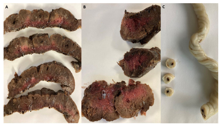
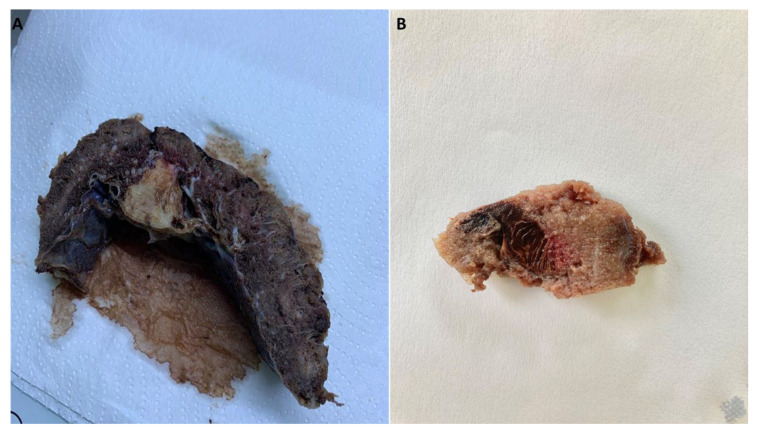
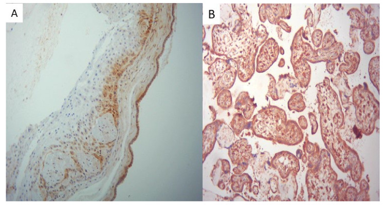
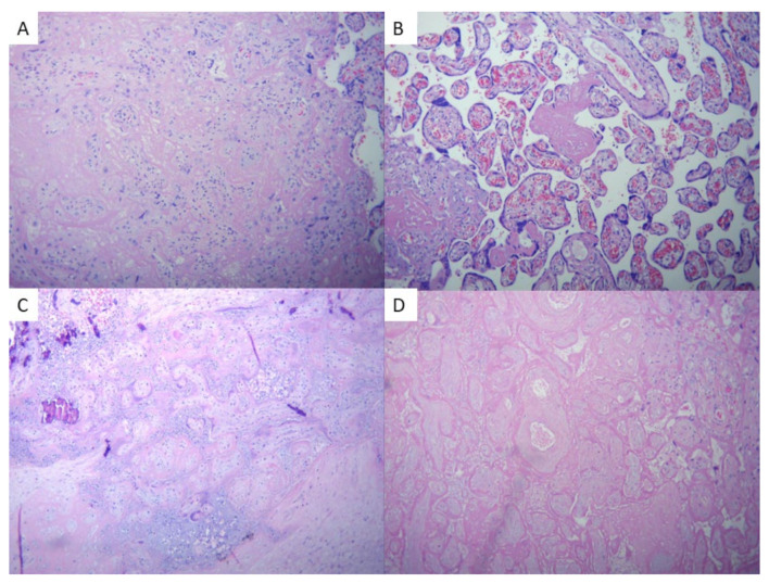
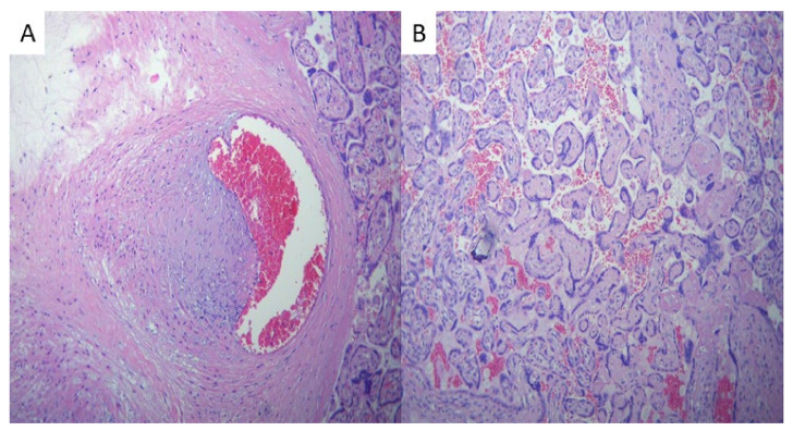
Research indicates compelling evidence of SARS-CoV-2 vertical transmission as a result of placental pathology. This study offers an approach to histopathological and immunohistochemical placental observations from SARS-CoV-2-positive mothers compared to negative ones. Out of the 44 examined placentas, 24 were collected from patients with a SARS-CoV-2 infection during pregnancy and 20 were collected from patients without infection. The disease group showed strong SARS-CoV-2 positivity of the membranes, trophoblasts, and fetal villous macrophages. Most infections occurred during the third trimester of pregnancy (66.6%). Pathology revealed areas consistent with avascular villi (AV) and thrombi in the chorionic vessels and umbilical cord in the positive group, suggesting fetal vascular malperfusion (FVM). This study shows SARS-CoV-2 has an impact on coagulation, demonstrated by fetal thrombotic vasculopathy (p = 0.01) and fibrin deposition (p = 0.01). Other observed features included infarction (17%), perivillous fibrin deposition (29%), intervillous fibrin (25%), delayed placental maturation (8.3%), chorangiosis (13%), chorioamnionitis (8.3%), and meconium (21%). The negative control group revealed only one case of placental infarction (5%), intervillous fibrin (5%), delayed placental maturation (5%), and chorioamnionitis (5%) and two cases of meconium (19%). Our study sheds light on the changes and differences that occurred in placentas from SARS-CoV-2-infected mothers and the control group. Further research is necessary to definitively establish whether SARS-CoV-2 is the primary culprit behind these intricate complications.
Keywords: COVID-19; SARS-CoV-2 infection; fetal; immunohistochemistry; pathology; placenta; pregnancy; vascular complications; vertical transmission.
Conflict of interest statement
The authors declare no conflicts of interest.
Figures
Similar articles
-
Histologic and Immunohistochemical Evaluation of 65 Placentas From Women With Polymerase Chain Reaction-Proven Severe Acute Respiratory Syndrome Coronavirus 2 (SARS-CoV-2) Infection.Arch Pathol Lab Med. 2021 Jun 1;145(6):648-656. doi: 10.5858/arpa.2020-0793-SA. Arch Pathol Lab Med. 2021. PMID: 33596304
-
Association Between COVID-19 Pregnant Women Symptoms Severity and Placental Morphologic Features.Front Immunol. 2021 May 26;12:685919. doi: 10.3389/fimmu.2021.685919. eCollection 2021. Front Immunol. 2021. PMID: 34122449 Free PMC article.
-
Extensive Perivillous Fibrin and Intervillous Histiocytosis in a SARS-CoV-2 Infected Placenta From an Uninfected Newborn: A Case Report Including Immunohistochemical Profiling.Pediatr Dev Pathol. 2021 Nov-Dec;24(6):581-584. doi: 10.1177/10935266211025122. Epub 2021 Jun 28. Pediatr Dev Pathol. 2021. PMID: 34176361
-
A structured review of placental morphology and histopathological lesions associated with SARS-CoV-2 infection.Placenta. 2020 Nov;101:13-29. doi: 10.1016/j.placenta.2020.08.018. Epub 2020 Aug 23. Placenta. 2020. PMID: 32911234 Free PMC article. Review.
-
Chronic Inflammatory Placental Disorders Associated With Recurrent Adverse Pregnancy Outcome.Front Immunol. 2022 Apr 22;13:825075. doi: 10.3389/fimmu.2022.825075. eCollection 2022. Front Immunol. 2022. PMID: 35529853 Free PMC article. Review.
Cited by
-
Risk Factors for Prematurity and Congenital Malformations in Assisted Reproductive Technology Pregnancies-A Retrospective Study.J Clin Med. 2024 Oct 29;13(21):6470. doi: 10.3390/jcm13216470. J Clin Med. 2024. PMID: 39518609 Free PMC article.
-
Extreme Prematurity: A Case Report on the Importance of Multidisciplinary Consultations Before and After Maternity Ward Discharge.Cureus. 2024 Dec 28;16(12):e76518. doi: 10.7759/cureus.76518. eCollection 2024 Dec. Cureus. 2024. PMID: 39872582 Free PMC article.
References
-
- Levitan D., London V., McLaren R.A., Jr., Mann J.D., Cheng K., Silver M., Balhotra K.S., McCalla S., Loukeris K. Histologic and Immunohistochemical Evaluation of 65 Placentas From Women With Polymerase Chain Reaction–Proven Severe Acute Respiratory Syndrome Coronavirus 2 (SARS-CoV-2) Infection. Arch. Pathol. Lab. Med. 2021;145:648–656. doi: 10.5858/arpa.2020-0793-SA. - DOI - PubMed
-
- Schwartz D.A., Bugatti M., Santoro A., Facchetti F. Molecular Pathology Demonstration of SARS-CoV-2 in Cytotrophoblast from Placental Tissue with Chronic Histiocytic Intervillositis, Trophoblast Necrosis and COVID-19. [(accessed on 6 November 2023)];J. Dev. Biol. 2021 9:33. doi: 10.3390/jdb9030033. Available online: https://www.mdpi.com/2221-3759/9/3/33. - DOI - PMC - PubMed
-
- Zaigham M., Gisselsson D., Sand A., Wikström A.-K., von Wowern E., Schwartz D.A., Iorizzo L., Nelander M., Blomberg M., Papadogiannakis N., et al. Clinical-Pathological Features in Placentas of Pregnancies with SARS-CoV-2 Infection and Adverse Outcome: Case Series with and without Congenital Transmission. BJOG Int. J. Obstet. Gynaecol. 2022;129:1361–1374. doi: 10.1111/1471-0528.17132. - DOI - PMC - PubMed
MeSH terms
Substances
LinkOut - more resources
Full Text Sources
Medical
Miscellaneous
