. 2024 Feb 2;25(3):1839.
doi: 10.3390/ijms25031839.

AAV-RPGR Gene Therapy Rescues Opsin Mislocalisation in a Human Retinal Organoid Model of RPGR-Associated X-Linked Retinitis Pigmentosa

Affiliations

AAV-RPGR Gene Therapy Rescues Opsin Mislocalisation in a Human Retinal Organoid Model of RPGR-Associated X-Linked Retinitis Pigmentosa

Paul E Sladen et al. Int J Mol Sci. .

Abstract

Variants within the Retinitis Pigmentosa GTPase regulator (RPGR) gene are the predominant cause of X-Linked Retinitis Pigmentosa (XLRP), a common and severe form of inherited retinal disease. XLRP is characterised by the progressive degeneration and loss of photoreceptors, leading to visual loss and, ultimately, bilateral blindness. Unfortunately, there are no effective approved treatments for RPGR-associated XLRP. We sought to investigate the efficacy of RPGRORF15 gene supplementation using a clinically relevant construct in human RPGR-deficient retinal organoids (ROs). Isogenic RPGR knockout (KO)-induced pluripotent stem cells (IPSCs) were generated using established CRISPR/Cas9 gene editing methods targeting RPGR. RPGR-KO and isogenic wild-type IPSCs were differentiated into ROs and utilised to test the adeno associated virus (AAV) RPGR (AAV-RPGR) clinical vector construct. The transduction of RPGR-KO ROs using AAV-RPGR successfully restored RPGR mRNA and protein expression and localisation to the photoreceptor connecting cilium in rod and cone photoreceptors. Vector-derived RPGR demonstrated equivalent levels of glutamylation to WT ROs. In addition, treatment with AAV-RPGR restored rhodopsin localisation within RPGR-KO ROs, reducing mislocalisation to the photoreceptor outer nuclear layer. These data provide mechanistic insights into RPGRORF15 gene supplementation functional potency in human photoreceptor cells and support the previously reported Phase I/II trial positive results using this vector construct in patients with RPGR-associated XLRP, which is currently being tested in a Phase III clinical trial.

Keywords: CRISPR/Cas9; IPSC; RPGR; X-linked; adeno associated virus; gene therapy; retinal organoids; retinitis pigmentosa.


Conflict of interest statement

All authors are affiliated with MeiraGTx. P.E.S., A.N., T.A-I., T.V., S.L.B., S.N., A.F., A.L. and A.G. are employees of MeiraGTx. M.M. provides consultancy services to MeiraGTx. The authors have no other relevant affiliations or financial involvement with any organisation or entity with a financial interest in or financial conflict with the subject matter or materials discussed in the manuscript apart from those disclosed.

Figures

Figure 1
Figure 1
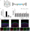
Assessment of RPGR-KO RO differentiation capacity. (A) WT and RPGR-KO d120 ROs express key retinal genes CRX, RECOVERIN, NRL and ARR3. Gene expression was first normalised to the geometric mean of GAPDH and ACTIN, then to d0 naive IPSCs. n = 1–4 ROs. (B) L/M Opsin (red) and Rhodopsin (RHO; white) expression in d160 WT and RPGR-KO ROs. Nuclei are identified with DAPI (blue). Zoomed panel denoted by dashed box. Scale bar = 50 μm.
Figure 2
Figure 2
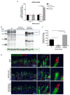
Characterisation of RPGR expression during retinal organoid development. (A,B) Analysis of RGPR isoform expression during RO development. RPGR isoform expression was first normalised to the geometric mean of GAPDH and ACTIN, then to d0 naive IPSCs. n = 1–4 ROs. (C) Immunoblotting of RPGR expression during RO development. RPGR1−19 (red box) is expressed at 140 kDa; RPGRORF15 (blue box) is detected at 200 kDa. Loading control β-tubulin (green box) is detected at 55 kDa. One organoid per lane. (D) Immunoblotting quantification demonstrates a significant reduction in RPGRORF15 protein in RPGR-KO ROs when compared to WT ROs. n = 2–4 ROs, ** p < 0.01, **** p < 0.0001. (E) RPGR119:RPGRORF15 ratio during WT RO differentiation.
Figure 3
Figure 3
Characterisation of AAV-RPGR treated ROs. (A) Schematic representation of AAV-RPGR. (B) Flow cytometry analysis of AAV capsid RO transduction efficiency. ROs were transduced with an eGFP transgene vector packaged in AAV7m8, AAV5 or AAV8. n = 4 ROs. (C) RPGR deficiency and AAV-RPGR treatment have no effect on retina-associated gene expression in d160 ROs. Gene expression was normalised to the geometric mean of GAPDH and ACTIN, and then to WT ROs. Red dashed line demarcates normalised WT expression levels. n = 3–6 ROs. (D) WT, non-transduced and AAV-RPGR transduced RPGR-KO d160 ROs express CRX (red) and RECOVERIN (REC; green). Nuclei are identified with DAPI (blue). Main image scale bar = 50 μm. Panel image scale bar = 20 μm.
Figure 4
Figure 4
AAV-RPGR restores RPGR expression and localisation in RPGR-KO ROs. (A) AAV-RPGR treatment significantly increases RPGRORF15 expression in RGPR-KO ROs. Gene expression was normalised to the geometric mean of GAPDH and ACTIN, and then to WT ROs. Red dashed line demarcates normalised WT expression levels. n = 3–6 ROs. **** p < 0.0001. (B) AAV-RPGR treatment restores RPGRORF15 (185 kDa, blue box) expression in d160 RPGR-KO ROs. Loading control β-tubulin (green box) is detected at 55 kDa. One organoid per lane. (C) Immunoblotting quantification demonstrates significantly increased RPGRORF15 protein levels in RPGR-KO ROs following AAV-RPGR transduction. n = 3–4 ROs. * p < 0.05, ** p < 0.01, *** p < 0.001. (D) RPGR localisation in the RO connecting cilium. WT ROs express RPGR protein (red) at the apical tip of the ciliary rootlet (ROOTLETIN; green). RPGR-KO ROs demonstrate mislocalised RPGR within the ciliary rootlet. AAV-RPGR transduction restores RPGR localisation in RPGR-KO ROs. Zoomed panels denoted by dashed boxes. Cell nuclei are identified with DAPI (blue). Arrows highlight the region of correct RPGR localisation. Main image scale bar = 10 μm, zoom image scale bar = 2 μm.
Figure 5
Figure 5
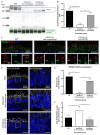
Restoration of RPGR glutamylation and rhodopsin localisation in RPGR-KO ROs by AAV-RPGR. (A) Immunoblotting analysis of RPGR glutamylation (GT335, blue box) in WT, non-transduced and AAV-RPGR-treated RPGR-KO organoids. Glutamylated β-tubulin (green box) is detected at 55 kDa. One organoid per lane. (B) Quantification of immunoblotting RPGR glutamylation levels, calculated as RPGR/β-tubulin ratio, demonstrates increased RPGR glutamylation in AAV-RPGR-treated RPGR-KO ROs compared to non-transduced ROs. n = 3–6 ROs, ** p < 0.01, *** p < 0.001. (C) ICC analysis of RGPR:GT335 co-localisation in d160 WT, non-transduced RPGR-KO and AAV-RPGR-treated RPGR-KO ROs. Arrows highlight areas of co-localisation between GT335 and RPGR in WT and AAV-RPGR treated ROs. Zoomed panel denoted by dashed box. Main image scale bar = 20 μm, zoom image scale bar = 2 μm. (D) ICC analysis of rhodopsin (white) mislocalisation in d160 WT, non-transduced RPGR-KO and AAV-RPGR-treated RPGR-KO ROs. Nuclei identified using DAPI (blue). Retina outer nuclear layer (ONL) denoted by red arrows. Zoomed panel denoted by dashed box. Scale bar = 40 μm, zoom scale bar = 10 μm. (E) Quantification of RPGR:GT335 co-localisation. n = 2–3 ROs. *** p < 0.001, **** p < 0.0001. (F) Quantification of rhodopsin mislocalisation, calculated as percentage of pixels above threshold in the ONL compared to total rhodopsin pixels. n = 2–3 ROs. * p < 0.05, ** p < 0.01.

References

    1. Awadh Hashem S., Georgiou M., Ali R.R., Michaelides M. RPGR-Related Retinopathy: Clinical Features, Molecular Genetics, and Gene Replacement Therapy. Cold Spring Harb. Perspect. Med. 2023;13:a041280. doi: 10.1101/cshperspect.a041280. - DOI - PMC - PubMed
    1. Lam B.L., Scholl H.P.N., Doub D., Sperling M., Hashim M., Li N. A Systematic Literature Review of Disease Progression Reported in RPGR-Associated X-Linked Retinitis Pigmentosa. Retina. 2023;44:1–9. doi: 10.1097/IAE.0000000000003920. - DOI - PubMed
    1. Meindl A., Dry K., Herrmann K., Manson F., Ciccodicola A., Edgar A., Carvalho M.R.S., Achatz H., Hellebrand H., Lennon A., et al. A Gene (RPGR) with Homology to the RCC1 Guanine Nucleotide Exchange Factor Is Mutated in X-Linked Retinitis Pigmentosa (RP3) Nat. Genet. 1996;13:35–42. doi: 10.1038/ng0596-35. - DOI - PubMed
    1. Jin Z.B., Liu X.Q., Hayakawa M., Murakami A., Nao-i N. Mutational Analysis of RPGR and RP2 Genes in Japanese Patients with Retinitis Pigmentosa: Identification of Four Mutations. Mol. Vis. 2006;12:1167–1174. - PubMed
    1. Vervoort R., Lennon A., Bird A.C., Tulloch B., Axton R., Miano M.G., Meindl A., Meitinger T., Ciccodicola A., Wright A.F. Mutational Hot Spot within a New RPGR Exon in X-Linked Retinitis Pigmentosa. Nat. Genet. 2000;25:462–466. doi: 10.1038/78182. - DOI - PubMed