. 2024 Feb 2;16(3):644.
doi: 10.3390/cancers16030644.

Keeping Pathologists in the Loop and an Adaptive F1-Score Threshold Method for Mitosis Detection in Canine Perivascular Wall Tumours

Affiliations

Keeping Pathologists in the Loop and an Adaptive F1-Score Threshold Method for Mitosis Detection in Canine Perivascular Wall Tumours

Taranpreet Rai et al. Cancers (Basel). .

Abstract

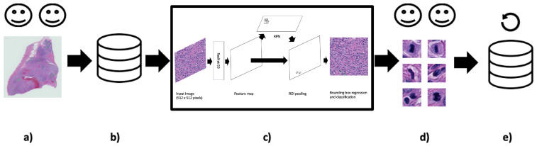
Performing a mitosis count (MC) is the diagnostic task of histologically grading canine Soft Tissue Sarcoma (cSTS). However, mitosis count is subject to inter- and intra-observer variability. Deep learning models can offer a standardisation in the process of MC used to histologically grade canine Soft Tissue Sarcomas. Subsequently, the focus of this study was mitosis detection in canine Perivascular Wall Tumours (cPWTs). Generating mitosis annotations is a long and arduous process open to inter-observer variability. Therefore, by keeping pathologists in the loop, a two-step annotation process was performed where a pre-trained Faster R-CNN model was trained on initial annotations provided by veterinary pathologists. The pathologists reviewed the output false positive mitosis candidates and determined whether these were overlooked candidates, thus updating the dataset. Faster R-CNN was then trained on this updated dataset. An optimal decision threshold was applied to maximise the F1-score predetermined using the validation set and produced our best F1-score of 0.75, which is competitive with the state of the art in the canine mitosis domain.

Keywords: artificial intelligence; canine Perivascular Wall Tumour; canine Soft Tissue Sarcoma; deep learning; digital pathology; faster R-CNN; humans in the loop; mitosis; mitosis detection; object detection; pathologists in the loop.


Conflict of interest statement

Zoetis funded part of the original study. The remaining authors declare that the research was conducted in the absence of any commercial or financial relationships that could be construed as a potential conflict of interest.

Figures

Figure A1
Figure A1
A depiction of the two-stage mitosis detection approach. On the top, in stage 1, 20× magnification images and annotations from the updated refined mitoses dataset are used to train a Faster R-CNN model (the model is also presented in Figure 1). Optimal probability thresholds are applied on the output candidates, which are determined from the validation set (based on Equation (4)). These selected candidates are then extracted (size 64 × 64 pixels) at 40× magnification from the original Whole Slide Images (WSIs) and passed into the second stage. On the bottom shows stage 2 where the extracted patches are fed into a DenseNet-161 ImageNet pre-trained feature extractor, where the outputs are fed into a logistic regression classifier to determine whether the candidates are mitosis or difficult false positives.
Figure A2
Figure A2
Receiver operating characteristic (ROC) curve plots from the second-stage logistic regression model results for each cross-validation fold. For each fold, it is evident that the models do not effectively learn the differences between true positive (TP) and false positive (FP) mitosis detections.
Figure 1
Figure 1
Image is inspired by Mahmood et al.’s depiction of Faster R-CNN [37]. A Faster R-CNN object detection model applied to the cPWT mitosis dataset. An input image of size 512 × 512 pixels is passed through the model where the feature map is extracted using the Resnet-50 feature-extraction network. This is then followed by generating region proposals in the Region Proposal Network (RPN) and finally mitosis detection in the classifier.
Figure 2
Figure 2
Keeping humans in the loop: (a) Two pathologist annotators independently review canine Perivascular Wall Tumour (cPWT) Whole Slide Images (WSIs) and applied centroid annotations to mitotic figures. (b) After initial agreement of mitoses, this formed the initial dataset of patch images with annotations. (c) A Faster R-CNN object detector was trained and tested on these data. (d) Thereafter, false positive (FP) candidates are reviewed again by the pathologist annotators where misclassified candidates are reassigned as true positives (TPs). (e) These new TPs are added to the updated dataset. (20× magnification images).
Figure 3
Figure 3
We used 20× magnification images and annotations from the updated mitosis dataset to train the Faster R-CNN object detection model (details from the Faster R-CNN model are also shown in Figure 1). Optimal thresholds using Equation (4) were applied on the output candidates determined from the validation set.
Figure 4
Figure 4
Line graphs that show the sensitivity, precision and F1-score calculated for each probability threshold for the three validation folds. To determine the optimal probability threshold, we choose the threshold with the highest F1-score as determined via Equation (4). In the above plots, these are denoted as “best threshold”. For fold 1, this threshold was 0.96, for fold 2, it was 0.84, and for fold 3, it was 0.91.
Figure 5
Figure 5
An example 512 × 512 pixel image from the test set with a false negative (FN) shown in the red bounding box and a false positive (FP) detection shown in the yellow bounding box (32 × 32 pixels). The FP detection provides a probability confidence score of 5.3% and so would typically be dismissed as a mitosis candidate once the adaptive F1-score threshold is applied.

References

    1. Bostock D., Dye M. Prognosis after surgical excision of canine fibrous connective tissue sarcomas. Vet. Pathol. 1980;17:581–588. doi: 10.1177/030098588001700507. - DOI - PubMed
    1. Dernell W.S., Withrow S.J., Kuntz C.A., Powers B.E. Principles of treatment for soft tissue sarcoma. Clin. Tech. Small Anim. Pract. 1998;13:59–64. doi: 10.1016/S1096-2867(98)80029-7. - DOI - PubMed
    1. Ehrhart N. Soft-tissue sarcomas in dogs: A review. J. Am. Anim. Hosp. Assoc. 2005;41:241–246. doi: 10.5326/0410241. - DOI - PubMed
    1. Mayer M.N., LaRue S.M. Soft tissue sarcomas in dogs. Can. Vet. J. 2005;46:1048. - PMC - PubMed
    1. Cavalcanti E.B., Gorza L.L., de Sena B.V., Sossai B.G., Junior M.C., Flecher M.C., Marcolongo-Pereira C., dos Santos Horta R. Correlation of Clinical, Histopathological and Histomorphometric Features of Canine Soft Tissue Sarcomas. Braz. J. Vet. Pathol. 2021;14:151–158. doi: 10.24070/bjvp.1983-0246.v14i3p151-158. - DOI

LinkOut - more resources