Review
. 2024 Feb 4;16(3):664.
doi: 10.3390/cancers16030664.

Management of Cutaneous Squamous Cell Carcinoma of the Scalp: The Role of Imaging and Therapeutic Approaches

Affiliations
Review

Management of Cutaneous Squamous Cell Carcinoma of the Scalp: The Role of Imaging and Therapeutic Approaches

Júlia Verdaguer-Faja et al. Cancers (Basel). .

Abstract

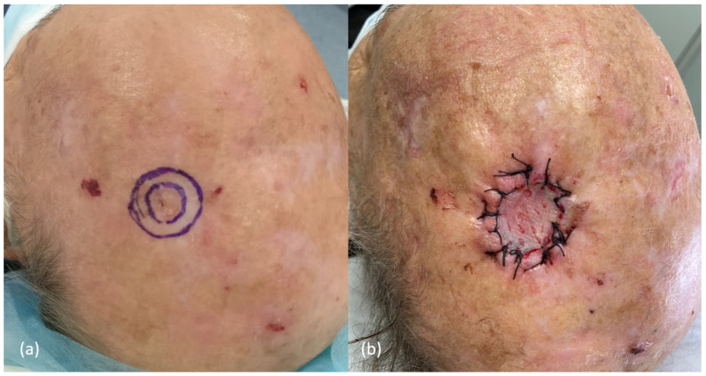
Cutaneous squamous cell carcinoma (cSCC) is the second most common subtype of skin cancer. The scalp is one of the most frequently affected locations and is associated with a higher rate of complications, compared to other locations. In addition, it has a characteristic thickness and anatomical structure that may influence both growth pattern and treatment of primary cSCC; while clinical peripheral margins may be easily achieved during the surgery, vertical excision of the tumor is limited by the skull. Despite having a unique anatomy, current guidelines do not contemplate specific recommendations for scalp cSCC, which leads to inconsistent decision-making in multidisciplinary committees when discussing tumors with high risk factors or with close margins. This article provides specific recommendations for the management of patients with scalp cSCC, based on current evidence, as well as those aspects in which evidence is lacking, pointing out possible future lines of research. Topics addressed include epidemiology, clinical presentation and diagnosis, imaging techniques, surgical and radiation treatments, systemic therapy for advanced cases, and follow-up. The primary focus of this review is on management of primary cSCC of the scalp with localized disease, although where relevant, some points about recurrent cSCCs or advanced disease cases are also discussed.

Keywords: cutaneous squamous cell carcinoma; head and neck; imaging; management; margins; recurrence; scalp; squamous cell carcinoma; surgery; treatment.


Conflict of interest statement

The authors declare no conflicts of interest.

Figures

Figure 1
Figure 1
Graphical representation of the anatomical structure of the scalp, with its five layers: epidermis + dermis, subcutaneous tissue, galea aponeurotica, loose areolar tissue, and periosteum. Blood vessels, lymphatics and nerves exist through the subcutaneous layer (small color circles), adjacent to fibrous tracts.
Figure 2
Figure 2
Clinical appearance of different cutaneous squamous cell carcinoma of the scalp. (a) Well-differentiated scalp cSCC. A rounded pink and hyperkeratotic tumor, with well-defined borders, in the parietal region of the scalp. (b) Moderately differentiated scalp cSCC. Hyperkeratotic erythematous plaque in the right parietal region, with poorly defined borders, and small areas with ulceration. Notice the actinic damage surrounding the lesion. (c) Poorly differentiated scalp cSCC. Erythematous and fleshy tumor, with a diameter greater than 2 cm, in the frontal region of an elderly patient.
Figure 3
Figure 3
Dermoscopy of different cutaneous squamous cell carcinomas. (a) Keratotic tumor, with yellowish-whitish keratosis in the center, with some hemorrhagic area, and a pink peripheral rim with hairpin and looped vessels. (b,c) Hyperkeratotic lesions with poorly defined edges, with an erythematous background with yellowish scales and keratosis, and small erosions. Few dotted vessels can be seen in the center of image (b).
Figure 4
Figure 4
Histological image of a cutaneous squamous cell carcinoma of the scalp, showing a proliferation of atypical squamous cells, ulcerated, that infiltrate the dermis, subcutaneous tissue, and galea aponeurotica (H&E, 0.6×).
Figure 5
Figure 5
Perineural invasion of a nerve of 0.05 mm in a cutaneous squamous cell carcinoma of the scalp (H&E, 63×).
Figure 6
Figure 6
(a,b) Clinical images of two cSCC satellitosis in the scalp, in an immunosuppressed patient, appearing as erythematous nodules next to the scar of a previously excised high-risk cSCC.
Figure 7
Figure 7
Proposed treatment algorithm for patients with primary cSCC of the scalp. cSCC: cutaneous squamous cell carcinoma; LN: lymph node; WLE: wide local excision; CT: computed tomography; CI: contraindication; US: ultrasound; RT: radiotherapy; SLNB: sentinel lymph node biopsy; MMS: Mohs micrographic surgery.
Figure 8
Figure 8
Example of the surgical approach performed in a cutaneous squamous cell carcinoma, <2 cm of diameter, in the left parietal region of an immunocompetent patient, using conventional surgery. (a) Peripheral clinical margins of 5 mm were performed. (b) As the histological margins were evaluated postoperatively, closure with a partial skin graft was chosen.
Figure 9
Figure 9
Proposed treatment algorithm for the management of histological margins and other histological features. RT: radiotherapy; cSCC: cutaneous squamous cell carcinoma; W&S: wait-and-see/close surveillance.
Figure 10
Figure 10
Proposed algorithm for the follow-up of cSCC of the scalp. cSCC: cutaneous squamous cell carcinoma; RLN: regional lymph node; CT: computed tomography. * However, the choice of imaging technique is left to the discretion of the clinician.

References

    1. Kwa R.E., Campana K., Moy R.L. Biology of Cutaneous Squamous Cell Carcinoma. J. Am. Acad. Dermatol. 1992;26:1–26. doi: 10.1016/0190-9622(92)70001-V. - DOI - PubMed
    1. Que S.K.T., Zwald F.O., Schmults C.D. Cutaneous Squamous Cell Carcinoma: Incidence, Risk Factors, Diagnosis, and Staging. J. Am. Acad. Dermatol. 2018;78:237–247. doi: 10.1016/j.jaad.2017.08.059. - DOI - PubMed
    1. Nagarajan P., Asgari M.M., Green A.C., Guhan S.M., Arron S.T., Proby C.M., Rollison D.E., Harwood C.A., Toland A.E. Keratinocyte Carcinomas: Current Concepts and Future Research Priorities. Clin. Cancer Res. 2019;25:2379–2391. doi: 10.1158/1078-0432.CCR-18-1122. - DOI - PMC - PubMed
    1. Andrade P., Vieira R., Reis J.P., Brites M.M., Mariano A. Epidemiology of Basal Cell Carcinomas and Squamous Cell Carcinomas in a Department of Dermatology–A 5 Year Review. An. Bras. Dermatol. 2012;87:212–219. doi: 10.1590/S0365-05962012000200004. - DOI - PubMed
    1. Youl P.H., Janda M., Aitken J.F., Del Mar C.B., Whiteman D.C., Baade P.D. Body-Site Distribution of Skin Cancer, Pre-Malignant and Common Benign Pigmented Lesions Excised in General Practice. Br. J. Dermatol. 2011;165:35–43. doi: 10.1111/j.1365-2133.2011.10337.x. - DOI - PubMed

LinkOut - more resources