. 2024 May 1;36(5):1806-1828.
doi: 10.1093/plcell/koae040.

Transcription factor PagMYB31 positively regulates cambium activity and negatively regulates xylem development in poplar

Affiliations

Transcription factor PagMYB31 positively regulates cambium activity and negatively regulates xylem development in poplar

Yanhui Zhang et al. Plant Cell. .

Abstract

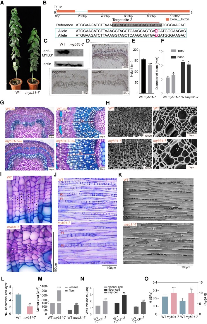
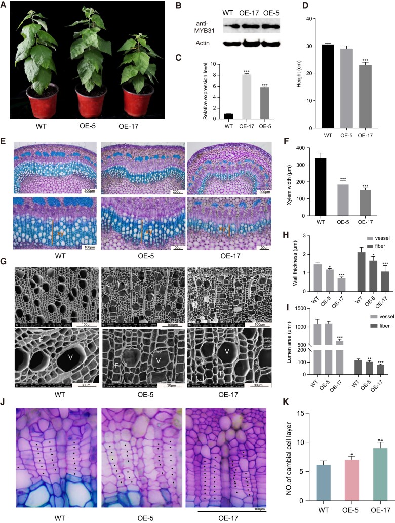
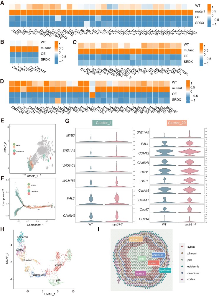
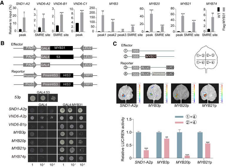
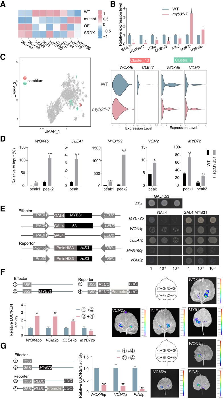
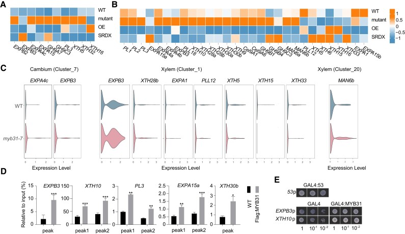
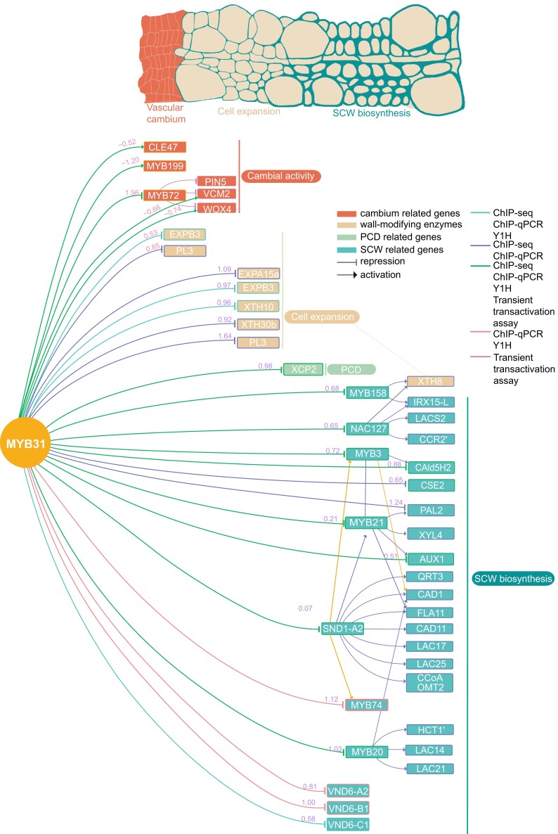
Wood formation involves consecutive developmental steps, including cell division of vascular cambium, xylem cell expansion, secondary cell wall (SCW) deposition, and programmed cell death. In this study, we identified PagMYB31 as a coordinator regulating these processes in Populus alba × Populus glandulosa and built a PagMYB31-mediated transcriptional regulatory network. PagMYB31 mutation caused fewer layers of cambial cells, larger fusiform initials, ray initials, vessels, fiber and ray cells, and enhanced xylem cell SCW thickening, showing that PagMYB31 positively regulates cambial cell proliferation and negatively regulates xylem cell expansion and SCW biosynthesis. PagMYB31 repressed xylem cell expansion and SCW thickening through directly inhibiting wall-modifying enzyme genes and the transcription factor genes that activate the whole SCW biosynthetic program, respectively. In cambium, PagMYB31 could promote cambial activity through TRACHEARY ELEMENT DIFFERENTIATION INHIBITORY FACTOR (TDIF)/PHLOEM INTERCALATED WITH XYLEM (PXY) signaling by directly regulating CLAVATA3/ESR-RELATED (CLE) genes, and it could also directly activate WUSCHEL HOMEOBOX RELATED4 (PagWOX4), forming a feedforward regulation. We also observed that PagMYB31 could either promote cell proliferation through the MYB31-MYB72-WOX4 module or inhibit cambial activity through the MYB31-MYB72-VASCULAR CAMBIUM-RELATED MADS2 (VCM2)/PIN-FORMED5 (PIN5) modules, suggesting its role in maintaining the homeostasis of vascular cambium. PagMYB31 could be a potential target to manipulate different developmental stages of wood formation.


Conflict of interest statement

Conflict of interest statement. The authors declare no competing interests.

Figures

Figure 1.
Figure 1.
Expression patterns of PagMYB31 in P. alba × P. glandulosa. A) RT-qPCR analysis of PagMYB31 expression in leaf (L), young root (R), differentiating xylem (X), and phloem–cambium (P-C). Values are the means ± Sd of 3 biological replicates. For comparison, the expression level in leaf is set as 1. B to F) GUS staining in tissue-cultured MYB31p-GUS transgenic P. alba × P. glandulosa.B) Young plant, C) cross section of stem with primary growth, D) enlarged image of vascular bundle, E) cross section of stem with secondary growth, and F) enlarged images of phloem, cambial zone, and xylem portions in E). F, fiber cell; P, pith; PF, phloem fiber; Ph, phloem; V, vessel; VC, vascular cambium; Xy, xylem.
Figure 2.
Figure 2.
PagMYB31 mutation decreases cambial activity and promotes cell expansion and SCW biosynthesis. A) An myb31 mutant and a WT plant. Plants are 5 mo old. B) Sequencing shows a base deletion at the target site 2 in the coding region of PagMYB31. C) Western blotting did not detect PagMYB31 proteins in the differentiating xylem of the myb31-7 mutant. D) Cellular immunolocalization of PagMYB31 proteins in the stem cross section of myb31-7 and WT plants. Cross section without anti-PagMYB31 antibody was used as a negative control. E) Height of 5-mo-old plants. F) Stem diameter in 7-mo-old plants. G) Cytological observations of stem cross sections of 5th and 10th internodes show enhanced xylem development. H) SEM of stem cross sections of 12th internode. I) Enlarged images in the cambial region of 10th internode. J) Cytological observations of tangential sections in the cambial region. K) SEM observation of tangential sections in the cambial region. L) The number of cambial cell layers at 10th internode. There are fewer layers of cambial cells in the myb31-7 mutant. M) Lumen area of fiber cells and vessels. N) Wall thickness of vessels, fiber cells and ray cells. O) NI analysis. In E), F), and L), error bars represent SD of 6 independent measurements. In M) and N), error bars represent Sd of 50 cells from 3 plants for each line. In O), error bars represent Sd of 20 points from 3 fiber cells in WT and myb31-7 plants. Asterisks show significant differences by Student's t-test (*P < 0.05; **P < 0.01, ***P < 0.001). F, fiber cell; FI, fusiform initial; RI, ray initial; V, vessel; VC: vascular cambium; Xy, xylem.
Figure 3.
Figure 3.
OE of Flag:PagMYB31 in P. alba × P. glandulosa inhibited xylem development and SCW thickening. A) WT plants and PagMYB31-OE transgenic lines 5 and 17 (OE-5, and -17). Plants are 1 mo old. B) Western blotting detection of PagMYB31 proteins in differentiating xylem of OE-17, OE-5 and WT. Actin was used as the internal control. C) RT-qPCR analysis of PagMYB31 expression in differentiating xylem of WT, OE-5 and OE-17. D) Height of 1-mo-old plants. E, G) Cytological observation on stem cross sections of 10th internode under light microscopy (E) and of 12th internode under SEM (G) in WT, OE-5 and OE-17. F, H, I) Measurement of xylem width in stem cross sections of 10th internode F), wall thickness of vessel and fibercells H) and lumen area of vessels and fiber cells I) of 12th internode. J) Enlarged images in the cambial region of WT, OE-5, and OE-17. K) Number of cambial cell layers. Error bars represent SD values of 3 biological replicates C), 6 independent measurements D, F, K) and 50 independent measurements H, I), and ANOVA was used for multiple-group comparison, followed by post hoc Dunnett pairwise comparisons to examine statistical significance between transgenic and WT plants (*P < 0.05; **P < 0.01; ***P < 0.001).
Figure 4.
Figure 4.
Transcriptome analysis of differentiating xylem in the myb31-7 mutant, Flag:MYB31, and MYB31:SRDX transgenic P. alba × P. glandulosa. Heat maps showing gene expression profiles of lignin biosynthetic pathway genes A), cellulose synthase (CesA) genes B), xylan biosynthesis enzyme genes C), and TF (MYB and NAC) genes D) in the differentiating xylem of WT, myb31-7 mutant, PagMYB31-OE and PagMYB31:SRDX OE (SRDX) transgenic plants. Orange and blue colors represent higher and lower gene expression levels. E) UMAP representation of clusters related to cambium and xylem by snRNA-seq. F) Pseudotime trajectory of cambium differentiation toward xylem. G) Violin plot visualization to compare the expression level in xylem Clusters 1 and 20 between WT and myb31-7 plants. The height of the violin represents the gene expression level, and the width of the violin represents the proportion of cells expressing in the cluster. H) UMAP visualization illustrates the cell clusters identified in spatial transcriptomics, with each color signifying a unique cell cluster. I) The cluster clusters are mapped over the images of hematoxylin–eosin staining section from 9th internode. 4CL, p-coumarate CoA ligase; C3H, p-coumaroyl-CoA 3-hydroxylase; C4H, cinnamate 4-hydroxylase; CAD, cinnamyl alcohol dehydrogenase; CAld5H, coniferaldehyde 5-hydroxylase; CCoAOMT, caffeoyl-CoA O-methyltransferase; CCR, cinnamoyl-CoA reductase; COMT, caffeic acid 3-O-methyltransferaese; CSE, caffeoyl shikimate esterase; HCT, hydroxycinnamoyltransferase; LAC, laccase; PAL, phenylalanine ammonia-lyase.
Figure 5.
Figure 5.
PagMYB31 regulates SCW biosynthesis through directly inhibiting the TF genes regulating SCW formation. A) Detection of the interactions between PagMYB31 and the promoters of 8 TF genes of SCW biosynthesis by ChIP-qPCR. ChIP was conducted in WT and Flag:PagMYB31-OE transgenic line OE-17. The qPCR primers are designed within the region of ChIP peaks, and SMRE sites were identified in the promoters of PagVND6-A2, B1, C1, MYB3/20, 21, and 74. B) Y1H assays of PagMYB31 with its targets. Cotransformants (GAL4:MYB31 and Promoter:HIS3) were diluted (1, 10−1, and 10−2) and grown on SD/-Leu-Trp-His medium, supplemented with a certain concentration of 3-AT. GLA4:53 + 53p:HIS3 was used as a positive control, and GAL4 + Promoter:HIS3 was the negative control. C) Effector–reporter-based activation/repression assays show that PagMYB31 inhibits the promoter activity of PagSND1-A2, MYB21 and 20/3. The activity of Rluc was used as internal interference. In A) and C), error bars represent Sd values of 3 biological replicates, and asterisks indicate significance between transgenic and WT plants by Student's t-test (*P < 0.05; **P < 0.01; ***P < 0.001).
Figure 6.
Figure 6.
PagMYB31 directly regulates the expression of the genes involved in cambial proliferation. A) Heat map shows the expression level changes of the genes involved in cambial cell proliferation in the phloem–cambium of WT, myb31-7 mutant, PagMYB31-OE and PagMYB31:SRDX OE (SRDX) transgenic plants. B) RT-qPCR confirmation of downregulation of PagWOX4, VMC2, MYB199, and PIN5 and upregulation of PagMYB72 and 196 in the phloem–cambium of the myb31-7 mutant. Specific primers are designed to quantify the transcript abundance of PagWOX4a + 4b and PagWOX4b. C) UMAP visualization (left) of Clusters 7 and 13, which represent cambium cells, and violin plot visualization (right) of the downregulation of PagWOX4, CLE47, and VCM2 in cambium clusters of the myb31-7 mutant by snRNA-seq. The height of the violin represents the gene expression level, and the width of the violin represents the proportion of cells expressing in the cluster. D) ChIP-qPCR shows the binding of PagMYB31 to the promoters of PagWOX4, CLE47, MYB199, VCM2, and MYB72. E) Y1H assays of PagMYB31 and the promoters of PagWOX4, CLE47, MYB199, VCM2, and MYB72. Cotransformants (GAL4:MYB31 and Promoter:HIS3) were diluted (1, 10−1, and 10−2) and grown on SD/-Leu-Trp-His medium, supplemented with 3-AT. GLA4:53 + 53p:HIS3 was used a positive control, and GAL4 + Promoter:HIS3 was the negative control. F) Effector–reporter assays show that PagMYB31 activates the promoter activity of PagWOX4, VCM2, and CLE47 but inhibits the promoter activity of PagMYB72. G) Effector–reporter transactivation/repression assays show that PagMYB72 represses the promoter activity of PagWOX4b, VCM2 and PIN5. In B), D), F), and G), error bars represent Sd values of 3 biological replicates, and asterisks indicate significance between transgenic and WT plants by Student's t-test (**P < 0.01; ***P < 0.001).
Figure 7.
Figure 7.
PagMYB31 regulates cambium and xylem cell expansion. A) Heat map shows the expression of wall-modifying enzyme genes, including expansin (EXPA) genes, XTH genes, and PL genes in the phloem–cambium A) and differentiating xylem B). C) Violin plot visualization to compare the expression level of wall-modifying enzyme genes in cambium and xylem clusters between WT and myb31-7 plants by snRNA-Seq. The height of the violin represents the gene expression level, and the width of the violin represents the proportion of cells expressing in the cluster. D) ChIP-qPCR detection of the binding of PagMYB31 to the promoters of 5 wall-modifying enzyme genes. Error bars represent Sd values of 3 biological replicates, and asterisks indicate significance between transgenic and WT plants by Student's t test (*P < 0.05; **P < 0.01; ***P < 0.001). E) Y1H assays of PagMYB31 with the promoters of PagEXPB3 and PagXTH10. Cotransformants (GAL4:MYB31 and Promoter:HIS3) were diluted (1, 10−1, and 10−2) and grown SD/-Leu-Trp-His, supplemented with 3-AT. GLA4:53 + 53p:HIS3 was used as the positive control, and GAL4 + Promoter:HIS3 was the negative control.
Figure 8.
Figure 8.
PagMYB31-mediated TRN regulating different developmental steps of wood formation, including cambial cell proliferation, xylem cell expansion, and SCW biosynthesis. In cambium, PagMYB31 directly regulates PagWOX4, VCM2, MYB199, and CLE47. PagMYB72 is the target of PagMYB31, and PagMYB72 directly inhibits PagWOX4, PagVCM2, and PagPIN5. PagMYB31 also inhibits the expression of wall-modifying enzyme genes (PagEXPB3 and PL3) to repress cambial activity. During xylem development, PagMYB31 directly inhibits the wall-modifying enzyme genes (PagEXPB3, EXP15a, XTH10, XTH30b, and PL3) to repress cell expansion. PagMYB31 can bind to the promoters of TF genes regulating SCW biosynthesis (4 SND1s, 6 VNDs, MYB3/20, 2/21, and 74), but only PagSND1-A2, MYB3/20, 74, PagVND6-A2, B1, and C1 were upregulated in the differentiating xylem of the myb31-7 mutant. The numbers in pink and purple colors on the lines represent the log2 fold change of PagMYB31 direct target genes in the cambium and differentiating xylem, respectively, of the myb31-7 mutant.

Similar articles

Cited by

References

    1. Avci U, Earl Petzold H, Ismail IO, Beers EP, Haigler CH. Cysteine proteases XCP1 and XCP2 aid micro-autolysis within the intact central vacuole during xylogenesis in Arabidopsis roots. Plant J. 2008:56(2):303–315. 10.1111/j.1365-313X.2008.03592.x - DOI - PubMed
    1. Berardini TZ, Reiser L, Li D, Mezheritsky Y, Muller R, Strait E, Huala E. The Arabidopsis information resource: making and mining the “gold standard” annotated reference plant genome. Genesis. 2015:53(8):474–485. 10.1002/dvg.22877 - DOI - PMC - PubMed
    1. Berthet S, Demont-Caulet N, Pollet B, Bidzinski P, Cézard L, Le Bris P, Borrega N, Hervé J, Blondet E, Balzergue S, et al. . Disruption of LACCASE4 and 17 results in tissue-specific alterations to lignification of Arabidopsis thaliana stems. Plant Cell. 2011:23(3):1124–1137. 10.1105/tpc.110.082792 - DOI - PMC - PubMed
    1. Brackmann K, Qi J, Gebert M, Jouannet V, Schlamp T, Grünwald K, Wallner ES, Novikova DD, Levitsky VG, Agustí J, et al. . Spatial specificity of auxin responses coordinates wood formation. Nat Commun. 2018:9(1):875. 10.1038/s41467-018-03256-2 - DOI - PMC - PubMed
    1. Butler A, Hoffman P, Smibert P, Papalexi E, Satija R. Integrating single-cell transcriptomic data across different conditions, technologies, and species. Nat Biotechnol. 2018:36(5):411–420. 10.1038/nbt.4096 - DOI - PMC - PubMed

Publication types

MeSH terms