. 2024 Feb 20;5(2):101418.
doi: 10.1016/j.xcrm.2024.101418. Epub 2024 Feb 9.

An enhanced broad-spectrum peptide inhibits Omicron variants in vivo

Affiliations

An enhanced broad-spectrum peptide inhibits Omicron variants in vivo

Wenwen Bi et al. Cell Rep Med. .

Abstract

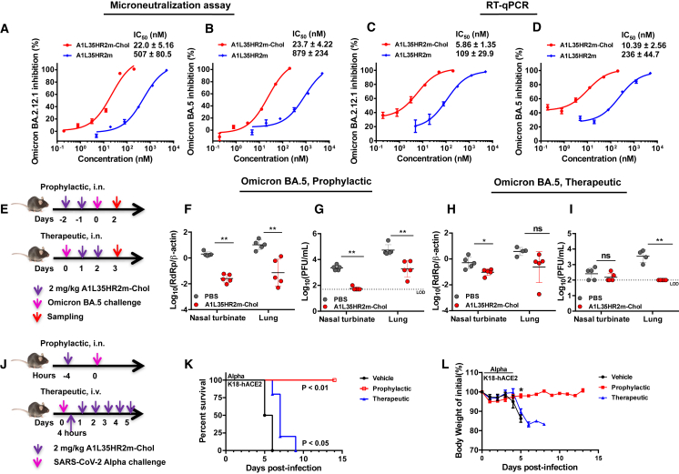
The continual emergence of severe acute respiratory syndrome coronavirus 2 (SARS-CoV-2) variants of concern (VOCs) poses a major challenge to vaccines and antiviral therapeutics due to their extensive evasion of immunity. Aiming to develop potent and broad-spectrum anticoronavirus inhibitors, we generated A1-(GGGGS)7-HR2m (A1L35HR2m) by introducing an angiotensin-converting enzyme 2 (ACE2)-derived peptide A1 to the N terminus of the viral HR2-derived peptide HR2m through a long flexible linker, which showed significantly improved antiviral activity. Further cholesterol (Chol) modification at the C terminus of A1L35HR2m greatly enhanced the inhibitory activities against SARS-CoV-2, SARS-CoV-2 VOCs, SARS-CoV, and Middle East respiratory syndrome coronavirus (MERS-CoV) pseudoviruses, with IC50 values ranging from 0.16 to 5.53 nM. A1L35HR2m-Chol also potently inhibits spike-protein-mediated cell-cell fusion and the replication of authentic Omicron BA.2.12.1, BA.5, and EG.5.1. Importantly, A1L35HR2m-Chol distributed widely in respiratory tract tissue and had a long half-life (>10 h) in vivo. Intranasal administration of A1L35HR2m-Chol to K18-hACE2 transgenic mice potently inhibited Omicron BA.5 and EG.5.1 infection both prophylactically and therapeutically.

Keywords: ACE2 peptide; HR2 peptide; SARS-CoV-2; cholesterol modification; lipopeptide; pan-coronavirus fusion inhibitor; synergistic effect; variants of concern.


Conflict of interest statement

Declaration of interests B.D., W.B., S.Y., K.T., and G.C. are the inventors of a provisional patent filed by Westlake University and The University of Hong Kong.

Figures

None
Graphical abstract
Figure 1
Figure 1
The design and characterization of A1L35HR2m, HR2mL35A1, A1L5HR2m, and A1HR2m (A) Design of the A1L35HR2m, HR2mL35A1, A1L5HR2m, and A1HR2m peptides and the sequences of the A1 and HR2m peptides. (B and C) Characterizations of A1L35HR2m, HR2mL35A1, A1L5HR2m, and A1HR2m by SDS-PAGE (B) and liquid chromatography-mass spectrometry (LC-MS) (C).
Figure 2
Figure 2
Conjugating the ACE2-derived peptide A1 to the N terminus of the HR2m peptide significantly enhanced the inhibitory activity against SARS-CoV-2 infection (A) Inhibitory activity of A1L35HR2m, HR2mL35A1, A1L5HR2m, and A1HR2m against SARS-CoV-2 pseudovirus infection in Caco-2 cells. (B) Inhibitory activity of A1L35HR2m, HR2m, A1, and A1/HR2m mixtures against SARS-CoV-2 pseudovirus infection in Caco-2 cells. (C and D) A1L35HR2m, HR2m, and A1 inhibition of SARS-CoV-2 D614G S-mediated cell-cell fusion in Caco-2 cells (C) and Calu-3 cells (D). Each sample was tested in triplicate, and the data are presented as the mean ± SEM. Each experiment was repeated at least twice. (E) Binding affinity of A1L35HR2m to HR1 was determined by BLI. (F) The binding affinity of HR2m to HR1 was determined by BLI. The fitting curves were analyzed by ForteBio Data Analysis 12.0 software.
Figure 3
Figure 3
Secondary structures of A1, HR2m, and A1L35HR2m alone or in complex with HR1 (A–C) The secondary structures of A1 (A), HR2m (B), and A1L35HR2m (C) alone or in complex with HR1 were analyzed with CD. The experiments were performed twice, and representative data are shown. NA, not available.
Figure 4
Figure 4
A1L35HR2m-Chol broadly and potently inhibited pseudotyped coronavirus infection (A) The chemical structure of A1L35HR2m-Chol. (B–K) Inhibitory activity of A1L35HR2m-Chol and A1L35HR2m in pseudovirus infection assays against SARS-CoV-2 D614G (B), SARS-CoV-2 Alpha variant (C), SARS-CoV-2 Beta variant (D), SARS-CoV-2 Delta variant (E), SARS-CoV-2 Omicron BA.1 variant (F), SARS-CoV-2 Omicron XBB variant (G), SARS-CoV-2 Omicron BQ.1.1 variant (H), SARS-CoV (I), MERS-CoV (J), and VSV-G (K). Each sample was tested in triplicate, and the data are presented as the mean ± SEM. Each experiment was repeated at least twice.
Figure 5
Figure 5
Ex vivo anti-SARS-CoV-2 D614G activity and peptide concentration in plasma or tissue homogenate samples of the peptide-treated mice (A and C) Dilution fold of the plasma (A) or tissue homogenate (C) samples from mice intranasally treated with A1L35HR2m (5 mg/kg, n = 3) or A1L35HR2m-Chol (5 mg/kg, n = 3) that can achieve 50% SARS-CoV-2 D614G inhibition. (B and D) Estimated concentrations of functionally active peptides in the plasma (B) or tissue homogenate (D) samples. Each sample was tested in triplicate, and the data are presented as the mean ± SEM. Each experiment was repeated at least twice.
Figure 6
Figure 6
A1L35HR2m-Chol potently inhibited authentic SARS-CoV-2 in vitro and in vivo (A and B) A1L35HR2m and A1L35HR2m-Chol inhibition of authentic Omicron BA.2.12.1 (A) and BA.5 (B) in microneutralization assays. (C and D) A1L35HR2m and A1L35HR2m-Chol inhibition of authentic Omicron BA.2.12.1 (C) and BA.5 (D) in viral load reduction assays. The viral load was quantified by RT-qPCR. (E) Experiment design of the A1L35HR2m-Chol prophylactic and therapeutic efficacy study against Omicron BA.5. K18-hACE2 transgenic mice were challenged with Omicron BA.5 (10,000 PFUs) at day 0. In the prophylactic study, A1L35HR2m-Chol (2 mg/kg) was intranasally administered 2 days and 1 day before infection. In the therapeutic study, A1L35HR2m-Chol (2 mg/kg) was intranasally administered at days 1 and 2 post-virus infection. (F and G) In the prophylactic experiment, nasal turbinates and lungs were collected 2 days post-infection and subjected to viral load (F) and infectious viral titer (G) detection. (H and I) In the therapeutic experiment, nasal turbinates and lungs were collected 3 days post-infection and subjected to viral load (H) and infectious virus titer (I) detection. (J) Schematic diagram of the prophylactic and therapeutic experimental design to evaluate the efficacy of A1L35HR2m-Chol in protecting mice from SARS-CoV-2 Alpha lethal infection. (K) Survival rate monitoring of the mice treated with PBS or prophylactic or therapeutic A1L35HR2m-Chol treatment. (L) Daily body weights of the mice. (A–D) Each sample was tested in triplicate, and the data are presented as the mean ± SEM. Each experiment was repeated at least twice. (F–L) PBS, n = 5; A1L35HR2m-Chol, n = 5. The dashed lines in (G) and (I) represent the limit of detection (LOD). Unpaired non-parametric Mann-Whitney test (F–I and L) or log-rank test (K) was performed. ∗p < 0.05 and ∗∗p < 0.01. ns denotes no significance.
Figure 7
Figure 7
A1L35HR2m-Chol potently inhibited Omicron EG.5.1 in vitro and in vivo (A and B) A1L35HR2m-Chol inhibition of authentic Omicron EG.5.1 in microneutralization (A) and viral load reduction (B) assays. The viral load was quantified by RT-qPCR. Each sample was tested in triplicate, and the data are presented as the mean ± SEM. Each experiment was repeated at least twice. (C) Experiment design of the A1L35HR2m-Chol prophylactic and therapeutic efficacy studies against Omicron EG.5.1. K18-hACE2 transgenic mice were challenged with Omicron EG.5.1 at day 0. In the prophylactic study, A1L35HR2m-Chol (2 mg/kg) was intranasally administered 4 h before infection. In the therapeutic study, A1L35HR2m-Chol (2 mg/kg) was intranasally administered 4 and 28 h post-viral infection. (D) Tissue samples were collected 48 h after viral infection and subjected to infectious viral titer detection by plaque assay. PBS, n = 5; A1L35HR2m-Chol, n = 5. The dashed line represents the LOD. Unpaired non-parametric Mann-Whitney test was performed. ∗∗p < 0.01. ns denotes no significance.

References

    1. Kuzmina A., Khalaila Y., Voloshin O., Keren-Naus A., Boehm-Cohen L., Raviv Y., Shemer-Avni Y., Rosenberg E., Taube R. SARS-CoV-2 spike variants exhibit differential infectivity and neutralization resistance to convalescent or post-vaccination sera. Cell Host Microbe. 2021;29:522–528.e2. doi: 10.1016/j.chom.2021.03.008. - DOI - PMC - PubMed
    1. Hoffmann M., Arora P., Groß R., Seidel A., Hörnich B.F., Hahn A.S., Krüger N., Graichen L., Hofmann-Winkler H., Kempf A., et al. SARS-CoV-2 variants B.1.351 and P.1 escape from neutralizing antibodies. Cell. 2021;184:2384–2393.e12. doi: 10.1016/j.cell.2021.03.036. - DOI - PMC - PubMed
    1. Garcia-Beltran W.F., Lam E.C., St Denis K., Nitido A.D., Garcia Z.H., Hauser B.M., Feldman J., Pavlovic M.N., Gregory D.J., Poznansky M.C., et al. Multiple SARS-CoV-2 variants escape neutralization by vaccine-induced humoral immunity. Cell. 2021;184:2523. doi: 10.1016/j.cell.2021.04.006. - DOI - PMC - PubMed
    1. Guo Y., Han J., Zhang Y., He J., Yu W., Zhang X., Wu J., Zhang S., Kong Y., Guo Y., et al. SARS-CoV-2 Omicron Variant: Epidemiological Features, Biological Characteristics, and Clinical Significance. Front. Immunol. 2022;13 doi: 10.3389/fimmu.2022.877101. - DOI - PMC - PubMed
    1. Rössler A., Riepler L., Bante D., von Laer D., Kimpel J. SARS-CoV-2 Omicron Variant Neutralization in Serum from Vaccinated and Convalescent Persons. N. Engl. J. Med. 2022;386:698–700. doi: 10.1056/NEJMc2119236. - DOI - PMC - PubMed

Publication types