

Metabolic and molecular imaging in inflammatory arthritis
- PMID: 38341194
- PMCID: PMC10862311
- DOI: 10.1136/rmdopen-2023-003880
Metabolic and molecular imaging in inflammatory arthritis
Abstract
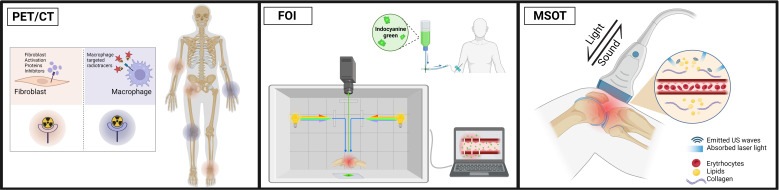
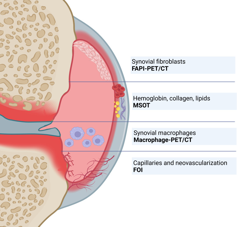
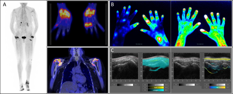
It is known that metabolic shifts and tissue remodelling precede the development of visible inflammation and structural organ damage in inflammatory rheumatic diseases such as the inflammatory arthritides. As such, visualising and measuring metabolic tissue activity could be useful to identify biomarkers of disease activity already in a very early phase. Recent advances in imaging have led to the development of so-called 'metabolic imaging' tools that can detect these changes in metabolism in an increasingly accurate manner and non-invasively.Nuclear imaging techniques such as 18F-D-glucose and fibroblast activation protein inhibitor-labelled positron emission tomography are increasingly used and have yielded impressing results in the visualisation (including whole-body staging) of inflammatory changes in both early and established arthritis. Furthermore, optical imaging-based bedside techniques such as multispectral optoacoustic tomography and fluorescence optical imaging are advancing our understanding of arthritis by identifying intra-articular metabolic changes that correlate with the onset of inflammation with high precision and without the need of ionising radiation.Metabolic imaging holds great potential for improving the management of patients with inflammatory arthritis by contributing to early disease interception and improving diagnostic accuracy, thereby paving the way for a more personalised approach to therapy strategies including preventive strategies. In this narrative review, we discuss state-of-the-art metabolic imaging methods used in the assessment of arthritis and inflammation, and we advocate for more extensive research endeavours to elucidate their full field of application in rheumatology.
Keywords: Arthritis; Arthritis, Rheumatoid; Inflammation; Lipids; Ultrasonography.
© Author(s) (or their employer(s)) 2024. Re-use permitted under CC BY-NC. No commercial re-use. See rights and permissions. Published by BMJ.
Conflict of interest statement
Competing interests: MW and FK are co-inventors, together with iThera Medical (Germany), on a European Union patent application (no. EP 19 163 304.9) relating to a device and a method for analysis of optoacoustic data, an optoacoustic system and a computer program. All other authors declare no conflicts of interest.
Figures
Similar articles
-
More advantages in detecting bone and soft tissue metastases from prostate cancer using 18F-PSMA PET/CT.Hell J Nucl Med. 2019 Jan-Apr;22(1):6-9. doi: 10.1967/s002449910952. Epub 2019 Mar 7. Hell J Nucl Med. 2019. PMID: 30843003
-
Imaging in arthritis: quantifying effects of therapeutic intervention using MRI and molecular imaging.Swiss Med Wkly. 2012 Jan 5;142:w13326. doi: 10.57187/smw.2012.13326. eCollection 2012. Swiss Med Wkly. 2012. PMID: 22252245
-
Imaging in inflammatory arthritis: progress towards precision medicine.Nat Rev Rheumatol. 2023 Oct;19(10):650-665. doi: 10.1038/s41584-023-01016-1. Epub 2023 Sep 8. Nat Rev Rheumatol. 2023. PMID: 37684361 Review.
-
Positron emission tomography/computed tomography imaging and rheumatoid arthritis.Int J Rheum Dis. 2014 Mar;17(3):248-55. doi: 10.1111/1756-185X.12316. Epub 2014 Mar 10. Int J Rheum Dis. 2014. PMID: 24606324 Review.
-
Diagnosis and management of rheumatoid arthritis; What is the current role of established and new imaging techniques in clinical practice?Best Pract Res Clin Rheumatol. 2016 Aug;30(4):586-607. doi: 10.1016/j.berh.2016.10.011. Epub 2016 Nov 22. Best Pract Res Clin Rheumatol. 2016. PMID: 27931956 Review.
Cited by
-
The Impact of IL-17A Inhibition in Rheumatic and Musculoskeletal Diseases: Current Insights and Future Prospects.Rheumatol Ther. 2025 Jun;12(3):435-451. doi: 10.1007/s40744-025-00754-w. Epub 2025 Apr 9. Rheumatol Ther. 2025. PMID: 40205297 Free PMC article. Review.
-
Unconventional Imaging Methods in Psoriatic Arthritis.Curr Rheumatol Rep. 2025 Jan 10;27(1):13. doi: 10.1007/s11926-024-01174-5. Curr Rheumatol Rep. 2025. PMID: 39792226 Free PMC article. Review.
-
Late-Onset Progressive Osseous Heteroplasia: 2 Unrelated Cases and Use of Positron Emission Tomography for Diagnosis.JCEM Case Rep. 2025 Feb 25;3(3):luae204. doi: 10.1210/jcemcr/luae204. eCollection 2025 Mar. JCEM Case Rep. 2025. PMID: 40008392 Free PMC article.
-
A method for quantifying and automatic grading of musculoskeletal ultrasound superb microvascular imaging based on dynamic analysis of optical flow model.Sci Rep. 2025 Apr 18;15(1):13369. doi: 10.1038/s41598-025-97924-1. Sci Rep. 2025. PMID: 40247053 Free PMC article.
-
Relationship between biologic therapy and cytokine levels in patients with inflammatory arthritis.Medicine (Baltimore). 2025 Jun 20;104(25):e42953. doi: 10.1097/MD.0000000000042953. Medicine (Baltimore). 2025. PMID: 40550053 Free PMC article.
References
-
- Just SA, Nielsen C, Werlinrud JC, et al. . Six-month prospective trial in early and long-standing rheumatoid arthritis: evaluating disease activity in the wrist through sequential synovial histopathological analysis, RAMRIS magnetic resonance score and EULAR-OMERACT ultrasound score. RMD Open 2019;5:e000951. 10.1136/rmdopen-2019-000951 - DOI - PMC - PubMed
Publication types
MeSH terms
LinkOut - more resources
Full Text Sources
Medical
Research Materials