. 2024;17(6):965-981.
doi: 10.1016/j.jcmgh.2024.01.023. Epub 2024 Feb 9.

Myeloid Deletion of Cdc42 Protects Liver From Hepatic Ischemia-Reperfusion Injury via Inhibiting Macrophage-Mediated Inflammation in Mice

Affiliations

Myeloid Deletion of Cdc42 Protects Liver From Hepatic Ischemia-Reperfusion Injury via Inhibiting Macrophage-Mediated Inflammation in Mice

Jing He et al. Cell Mol Gastroenterol Hepatol. 2024.

Abstract

Background & aims: Hepatic ischemia-reperfusion injury (HIRI) often occurs in liver surgery, such as partial hepatectomy and liver transplantation, in which myeloid macrophage-mediated inflammation plays a critical role. Cell division cycle 42 (Cdc42) regulates cell migration, cytoskeleton rearrangement, and cell polarity. In this study, we explore the role of myeloid Cdc42 in HIRI.

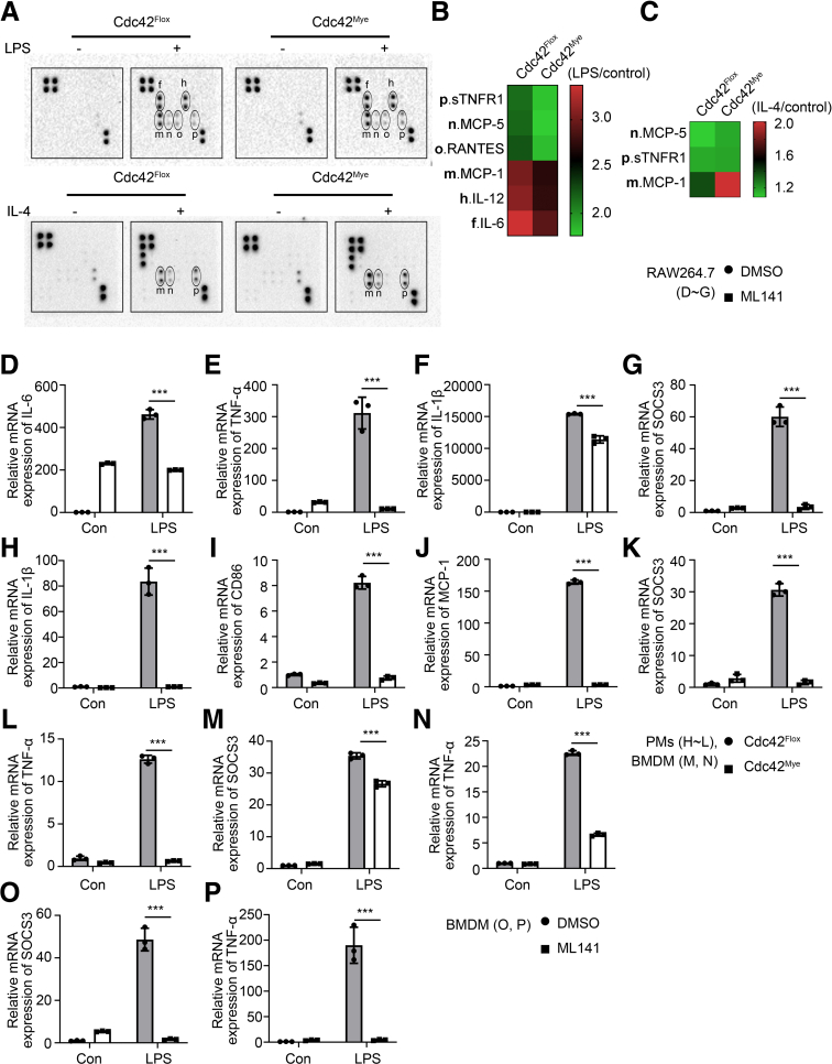
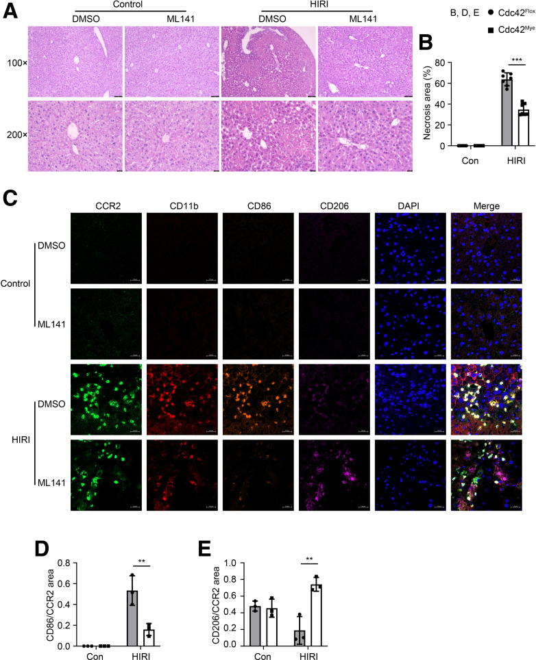
Methods: Mouse HIRI models were established with 1-hour ischemia followed by 12-hour reperfusion in myeloid Cdc42 knockout (Cdc42mye) and Cdc42flox mice. Myeloid-derived macrophages were traced with RosamTmG fluorescent reporter under LyzCre-mediated excision. The experiments for serum or hepatic enzymic activities, histologic and immunologic analysis, gene expressions, flow cytometry analysis, and cytokine antibody array were performed.

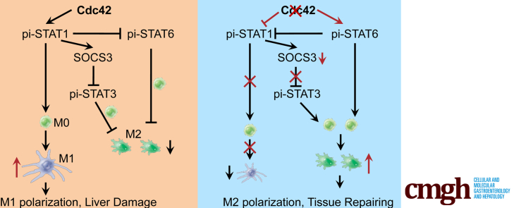
Results: Myeloid deletion of Cdc42 significantly alleviated hepatic damages with the reduction of hepatic necrosis and inflammation, and reserved hepatic functions following HIRI in mice. Myeloid Cdc42 deficiency suppressed the infiltration of myeloid macrophages, reduced the secretion of proinflammatory cytokines, restrained M1 polarization, and promoted M2 polarization of myeloid macrophages in livers. In addition, inactivation of Cdc42 promoted M2 polarization via suppressing the phosphorylation of STAT1 and promoting phosphorylation of STAT3 and STAT6 in myeloid macrophages. Furthermore, pretreatment with Cdc42 inhibitor, ML141, also protected mice from hepatic ischemia-reperfusion injury.

Conclusions: Inhibition or deletion of myeloid Cdc42 protects liver from HIRI via restraining the infiltration of myeloid macrophages, suppressing proinflammatory response, and promoting M2 polarization in macrophages.

Keywords: Cell Division Cycle 42 (Cdc42); Hepatic Ischemia-Reperfusion Injury; Macrophage; Polarization.


Figures

None
Graphical abstract
Figure 1
Figure 1
Myeloid deletion of Cdc42 alleviates the liver damages induced by HIRI in mice. Mouse HIRI model was established with 1-hour ischemia followed by 12-hour reperfusion in myeloid Cdc42 knockout (Cdc42mye) and wild-type (Cdc42flox) mice, N=12 per group. Serum alanine transaminase (ALT) (A) and aspartate aminotransferase (AST) (B) were measured in HIRI models from Cdc42mye and Cdc42flox mice, n=3-11 as indicated. Histologic images were taken from hematoxylin-eosin staining in mouse liver tissues with or without HIRI (C, D), scale bars = 50 μm, n=3. Apoptotic hepatocytes were determined by 1-step terminal-deoxynucleotidyl transferase mediated nick end labeling (TUNEL) analysis in liver tissues (E), n=3-5 as indicated. The lactate dehydrogenase (LDH) activities (F), malondialdehyde (MDA) contents (G), and superoxide dismutase (SOD) activities (H) were measured in liver tissues of mice, n=3-12 as indicated. The results were presented as mean ± standard deviation with at least 3 replicates. ∗P < .05; ∗∗P < .01; ∗∗∗P < .001.
Figure 2
Figure 2
Myeloid deletion of Cdc42 suppressed macrophage infiltration in HIRI liver tissues. Myeloid-derived macrophages are traced by fluorescence with Rosa mTmG reporter in liver tissues in HIRI. A schematic diagram for generation of myeloid Rosa mTmG/LyzCre mice or myeloid Cdc42mye/Rosa mTmG/LyzCre mice, in which LyzCre+ myeloid lineage cells were green fluorescent protein (GFP, mG) positive in green, other cells were expressing Tomato fluorescent proteins (mT) in red (A). Representative fluorescent images of livers from Rosa mTmG/LyzCre mice and Cdc42mye/Rosa mTmG/LyzCre mice were shown (B). The hepatic localization of CD11B-positive cells were labeled with immunofluorescence in sham and HIRI mice (C). scale bars = 50 μm, n = 3. ∗∗∗P < .001.
Figure 3
Figure 3
Cdc42 deficiency suppressed macrophage M1 polarization and promoted macrophage M2 polarization in HIRI livers in mice. An mIF experiment, based on tyramide signal amplification method was performed with CCR2, CD11B, CD86, and CD206 (A, C, E) or CCR2, CD11B, iNOS, and CD206 (B, D) on HIRI liver tissue sections in myeloid Cdc42 knockout (Cdc42mye) and wild-type (Cdc42flox) mice. scale bars = 20 μm, n=3, ∗∗P < 0.01; ∗∗∗P < 0.001.
Figure 4
Figure 4
Myeloid deletion of Cdc42 inhibits HIRI-induced inflammatory response in mice. Blood cell components such as WBC (A), red blood cell (B), lymph (C), granulocytes (Gran) (D), and Mid (E) were examined by routine blood analysis, n=7-8 as indicated. FCA was performed with CD11b, Ly6C or Ly6G labeling in blood cells (F–H), n=3-4 as indicated, and the mRNA expressions of TNF-α (I), IL1β (J), IL6 (K), and CD206 (L) were determined by qRT-PCR analysis in liver tissues, n=3-6 as indicated. Mouse serum cytokines (M, N) were detected with mouse cytokine antibody array in HIRI models from Cdc42mye compared with Cdc42flox mice. n. s., no significance; ∗P < .05; ∗∗P < .01; ∗∗∗P < .001.
Figure 5
Figure 5
Myeloid deletion of Cdc42 facilitates M2-type polarization of macrophages in HIRI model of mice. Expressions of CD206 were determined by immunohistochemical staining analysis with antibody against CD206 in liver tissues of Cdc42mye and Cdc42flox mice, scale bars = 50 μm or 20 μm as indicated, respectively (A), n=3-5 as indicated. The images (B) and quantitative results of IL6 (C), IL10 (D), iNOS (E), and ARG1 (F) were analyzed by Western blot assay in mouse liver tissues, n=4-9 as indicated. FCA of Ly6C, CD11b, CD86, and CD206 labeling in blood cells for M1/M2 macrophage typing were performed (G, H) ,n=3-5 as indicated. ∗∗P < .01; ∗∗∗P < .001.
Figure 6
Figure 6
Myeloid deletion of Cdc42 reduces the releases of inflammatory cytokines in macrophages in vitro. BMDMs were isolated from Cdc42mye and Cdc42flox mice. Mouse cytokines were determined by mouse cytokine antibody array with the culture medium of the mouse BMDMs stimulated with LPS (100 ng/mL) for 24 hours (A, B), or IL4 (40 ng/mL) for 48 hours (A, C), respectively. The mRNA expressions of IL6, TNF-α, IL1β, and SOCS3 were examined by qRT-PCR analysis in RAW264.7 cells, in which the cells were pretreated with ML141 (10 μM), a Cdc42 suppressor, for 2 hours, and then stimulated with LPS (100 ng/mL) for 6 hours (D–G), n=3. The mRNA expressions of IL1β, CD86, and MCP1 in mouse PMs (H–J) with LPS 100 ng/mL for 12 hours, n=3. The expressions of SOCS3 and TNF-α were detected by qRT-PCR with LPS (100 ng/mL) for 12 hours in mouse PMs (K, L) and BMDMs from myeloid Cdc42 knockout (Cdc42mye) and wild-type (Cdc42flox) mice (M, N), or BMDMs pretreated with 10 μM ML141 for 2 hours, and stimulated with LPS (100 ng/mL) for 12 hours, respectively (O, P). n=3. ∗∗∗P < .001.
Figure 7
Figure 7
Cdc42 suppression participated in macrophage polarization of THP1. Differentiation of THP1 cells were determined by FCA, in which the cells were labeled with antibodies of CD11b-APC/CD206-FITC, CD11b-APC/CD86-FITC, or CD11b-APC/CD64-FITC, respectively (A–C). THP1 cells were differentiated with PMA 100 ng/mL for 24 hours, then the cells were pretreated with 10 μM ML141 for 2 hours, and further stimulated with LPS (100 ng/mL) and IFN-γ (20 ng/mL) for 24 hours, or IL4 and IL13 (both 40 ng/mL) for 48 hours. n=3. ∗P < .05; ∗∗P < .01.
Figure 8
Figure 8
Myeloid deletion of Cdc42 enhances the M2-type differentiation of macrophages via regulating STATs signaling. THP1 cells were pretreated with PMA (100 ng/mL) and ML141 (10 μM) for differentiation and then stimulated with LPS (100 ng/mL) and IFN-γ (20 ng/mL) for 1 hour for M1 induction, or IL4 and IL13 (both at 40 ng/mL) for 48 hours for M2 induction. The images (A) and quantitative results (C–F) of the phosphorylation of STAT1, STAT3, and STAT6 and expressions of SOCS3 were detected by Western blot analysis in THP1 cells, n = 3. The images (B) and quantitative results (G–J) of the phosphorylation of STAT1, STAT3, and STAT6 and expressions of SOCS3 were measured by Western blot analysis in BMDMs stimulated with LPS (100 ng/mL) for 3 hours or IL4 (40 ng/mL) for 48 hours, respectively, n = 3. n. s., no significance; ∗P < .05; ∗∗∗P < .001.
Figure 9
Figure 9
Inhibition of Cdc42 with ML141 protects mice from HIRI. Cdc42 specific inhibitor ML141 was prepared in a sterilized injection solution (2% DMSO+30% PEG 300+5% Tween 80), and administered intraperitoneally to C57/BL6 mice at a dose of 10 mg/kg. Mouse HIRI model was performed 24 hours later as previously described. Hepatic histologic images were taken and analyzed from at least 3 mice per group with or without ML141 injection (A, B), scale bars at 75 or 25 μm, n=3. The mIF experiment, based on Tyramide signal amplification method (TSA) was performed with CCR2, CD11b, CD86, and CD206 on liver tissue sections (C), n=3. scale bars = 20 μm, ∗∗P < 0.01; ∗∗∗P < .001.

Similar articles

Cited by

References

    1. Saidi R.F., Kenari S.K. Liver ischemia/reperfusion injury: an overview. J Invest Surg. 2014;27:366–379. - PubMed
    1. Hirao H., Nakamura K., Kupiec-Weglinski J.W. Liver ischaemia-reperfusion injury: a new understanding of the role of innate immunity. Nat Rev Gastroenterol Hepatol. 2022;19:239–256. - PubMed
    1. Konishi T., Lentsch A.B. Hepatic ischemia/reperfusion: mechanisms of tissue injury, repair, and regeneration. Gene Expr. 2017;17:277–287. - PMC - PubMed
    1. Wen Y., Lambrecht J., Ju C., et al. Hepatic macrophages in liver homeostasis and diseases-diversity, plasticity and therapeutic opportunities. Cell Mol Immunol. 2021;18:45–56. - PMC - PubMed
    1. Liaskou E., Wilson D.V., Oo Y.H. Innate immune cells in liver inflammation. Mediators Inflamm. 2012;2012 - PMC - PubMed

MeSH terms