

The Role of Trabecular, Ligamentous-Intervertebral Disk and Facet Joints Systems: A Finite Element Analysis in the L4-S1 Vertebrae
- PMID: 38343310
- PMCID: PMC11571555
- DOI: 10.1177/21925682241231525
The Role of Trabecular, Ligamentous-Intervertebral Disk and Facet Joints Systems: A Finite Element Analysis in the L4-S1 Vertebrae
Abstract
Study design: Descriptive.
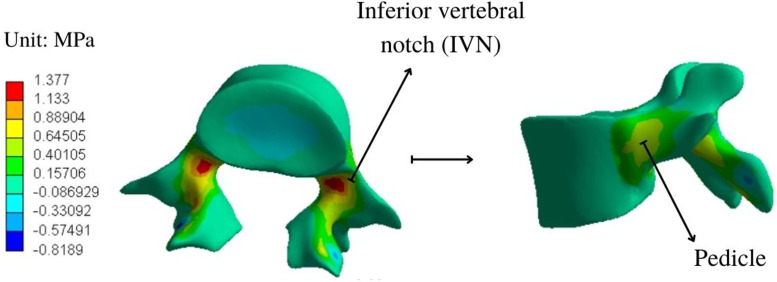
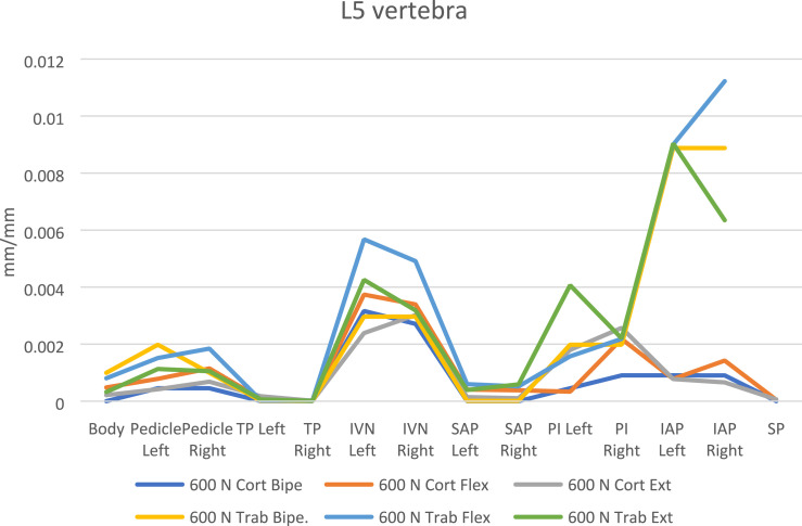
Objectives: Trabecular bone in the vertebrae is critical for the distribution of load and stress throughout the neuroaxis, as well as the intervertebral disk, ligamentous complex, and facet joints. The objective was to assess the stress and strain distribution of the L4-S1 spine segment by a finite element analysis.
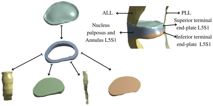
Methods: A lumbosacral spine model was built based on a CT-Scan. Trabecular-to-cortical bone distribution, ligaments, intervertebral disk, and facet joints with cartilage were included. A perpendicular force was applied over the L4 upper terminal plate of 300 N, 460 N and 600 N in neutral, plus 5 Nm and 7.5 Nm for flexion and extension movements. Maximum principal stress and total deformation were the main studied variables.
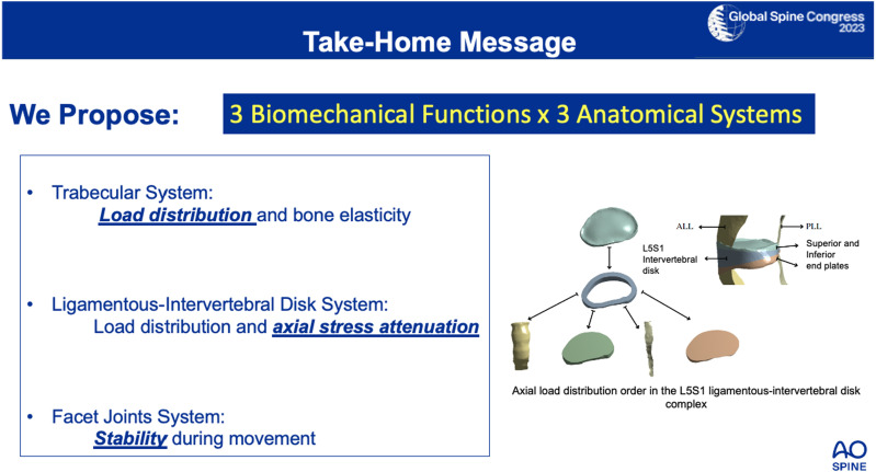
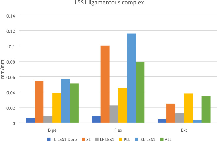
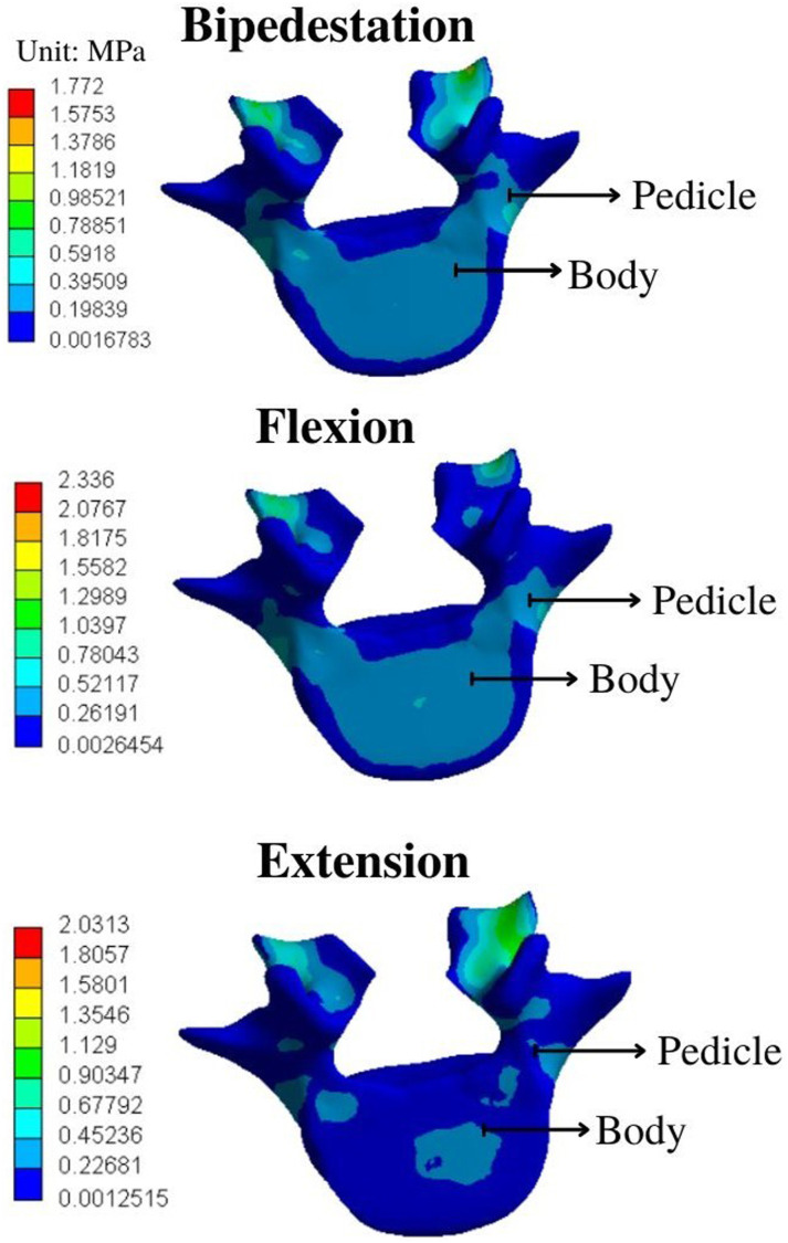
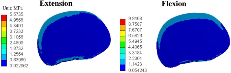
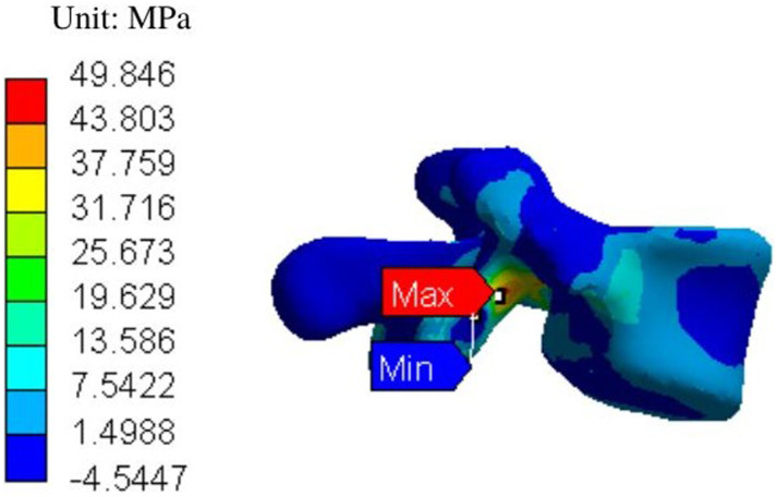
Results: Trabecular bone confers resistance to axial loads on the vertebrae by elastic capacity and stress distribution. MPS and TD showed axial stress attenuation in the nucleus pulposus and longitudinal ligaments, as well as load distribution capacity. Facet joints and discontinuous ligaments showed greater TD values in flexion moments but greater MPS values in extension, conferring stability to the lumbosacral junction and axial load distribution.
Conclusion: We propose 3 anatomical systems for axial load distribution and stress attenuation in the lumbosacral junction. Trabecular bone distributes loads, while the ligamentous-intervertebral disk transmits and attenuate axial stress. Facet joints and discontinuous ligaments act as stabilizers for flexion and extension postures. Overall, the relationship between trabecular bone, ligamentous-intervertebral disk complex and facet joints is necessary for an efficient load distribution and segmental axial stress reduction.This slide can be retrieved from the Global Spine Congress 2023.
Keywords: biomechanics; finite element analysis; lumbar vertebrae; lumbosacral region; trabecular bone.
Conflict of interest statement
Declaration of Conflicting InterestsThe author(s) declared the following potential conflicts of interest with respect to the research, authorship, and/or publication of this article: Brayan Felipe Pinzón received a 16-month contract with monthly paid salary by the ECCI University, as a Young researcher. Every autor listed was provided with access to software licenses and hardware acquisition for data processing by the Hospital Universitario de la Samaritana, used in this manuscript.
Figures


Similar articles
-
On the load-sharing along the ligamentous lumbosacral spine in flexed and extended postures: Finite element study.J Biomech. 2016 Apr 11;49(6):974-982. doi: 10.1016/j.jbiomech.2015.09.050. Epub 2015 Oct 20. J Biomech. 2016. PMID: 26493346
-
Analyzing isolated degeneration of lumbar facet joints: implications for degenerative instability and lumbar biomechanics using finite element analysis.Front Bioeng Biotechnol. 2024 Mar 27;12:1294658. doi: 10.3389/fbioe.2024.1294658. eCollection 2024. Front Bioeng Biotechnol. 2024. PMID: 38600941 Free PMC article.
-
Biomechanical Effect of L4 -L5 Intervertebral Disc Degeneration on the Lower Lumbar Spine: A Finite Element Study.Orthop Surg. 2020 Jun;12(3):917-930. doi: 10.1111/os.12703. Epub 2020 May 31. Orthop Surg. 2020. PMID: 32476282 Free PMC article.
-
Experimental measurement of ligament force, facet force, and segment motion in the human lumbar spine.J Biomech. 1993 Apr-May;26(4-5):427-38. doi: 10.1016/0021-9290(93)90006-z. J Biomech. 1993. PMID: 8478347 Review.
-
Spinal facet joint biomechanics and mechanotransduction in normal, injury and degenerative conditions.J Biomech Eng. 2011 Jul;133(7):071010. doi: 10.1115/1.4004493. J Biomech Eng. 2011. PMID: 21823749 Free PMC article. Review.
References
-
- Oliveira C, Navarro Garcia R, Ruiz Caballero J, Brito Ojeda E. Biomecánica de la columna vertebral. Canar Médica Quirúrgica. 2007;4:35-43.
-
- Sanabria MV. Anatomía y Exploración Física de la Columna Cervical y Torácica. 29. Costa Rica: Med Leg Costa Rica; 2012.
-
- Luque Sendra MI. Estudio de la Morfología del Cuerpo Verterbral en Una L4 Humana Con Modelos de Remodelación Ósea Interna y Externa [Pregrado]. [Sevilla]: Universidad de Sevilla; 2009.
-
- Drake R, Vogl W, Mitchell A, Gray H. Anatomía Para Estudiantes. 4.a ed. Barcelona: Elsevier; 2020.
LinkOut - more resources
Full Text Sources
