. 2024 Mar;103(3):318-328.
doi: 10.1177/00220345231222173. Epub 2024 Feb 12.

Murine IRF8 Mutation Offers New Insight into Osteoclast and Root Resorption

Affiliations

Murine IRF8 Mutation Offers New Insight into Osteoclast and Root Resorption

A Das et al. J Dent Res. 2024 Mar.

Abstract

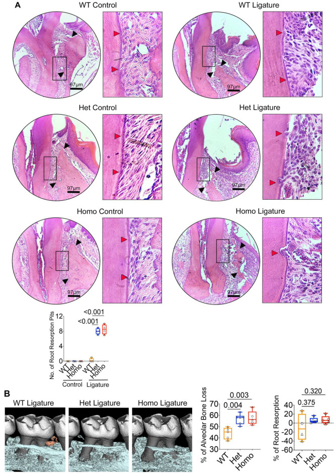
Interferon regulatory factor 8 (IRF8), a transcription factor expressed in immune cells, functions as a negative regulator of osteoclasts and helps maintain dental and skeletal homeostasis. Previously, we reported that a novel mutation in the IRF8 gene increases susceptibility to multiple idiopathic cervical root resorption (MICRR), a form of tooth root resorption mediated by increased osteoclast activity. The IRF8 G388S variant in the highly conserved C-terminal motif is predicted to alter the protein structure, likely impairing IRF8 function. To investigate the molecular basis of MICRR and IRF8 function in osteoclastogenesis, we generated Irf8 knock-in (KI) mice using CRISPR/Cas9 technique modeling the human IRF8G388S mutation. The heterozygous (Het) and homozygous (Homo) Irf8 KI mice showed no gross morphological defects, and the development of hematopoietic cells was unaffected and similar to wild-type (WT) mice. The Irf8 KI Het and Homo mice showed no difference in macrophage gene signatures important for antimicrobial defenses and inflammatory cytokine production. Consistent with the phenotype observed in MICRR patients, Irf8 KI Het and Homo mice demonstrated significantly increased osteoclast formation and resorption activity in vivo and in vitro when compared to WT mice. The oral ligature-inserted Het and Homo mice displayed significantly increased root resorption and osteoclast-mediated alveolar bone loss compared to WT mice. The increased osteoclastogenesis noted in KI mice is due to the inability of IRF8G388S mutation to inhibit NFATc1-dependent transcriptional activation and downstream osteoclast specific transcripts, as well as its impact on autophagy-related pathways of osteoclast differentiation. This translational study delineates the IRF8 domain important for osteoclast function and provides novel insights into the IRF8 mutation associated with MICRR. IRF8G388S mutation mainly affects osteoclastogenesis while sparing immune cell development and function. These insights extend beyond oral health and significantly advance our understanding of skeletal disorders mediated by increased osteoclast activity and IRF8's role in osteoclastogenesis.

Keywords: NFATC Transcription Factors; dental biology; genetic animal models; genetic mutation; human association studies; osteoclasts.


Conflict of interest statement

Declaration of Conflicting InterestsThe authors declared no potential conflicts of interest with respect to the research, authorship, and/or publication of this article.

Figures

Figure 1.
Figure 1.
Generation of Irf8 knock-in (KI) mice. (A) Schematic illustration of the G388S mutation in the mouse Irf8 gene. Displayed at the top is the structure of the mouse Irf8 gene, with introns represented by lines and the direction of transcription indicated by arrows. Exons are depicted as solid boxes, with coding regions as wide boxes and untranslated regions (UTRs) as narrow boxes. Directly beneath the gene structure is a diagram illustrating important domains of the IRF8 protein. The wild-type (WT) and mutant DNA sequences are listed at the bottom, with the CRISPR single-guide RNA (sgRNA) binding region underlined and the PAM shown in bold. The altered amino acid and its codon are shown in red, while a silent mutation is shown in blue. The silent mutation does not result in amino acid change, but it can facilitate the use of polymerase chain reaction (PCR) to differentiate the WT and KI alleles. (B) Sanger sequencing validated G388S knock-in allele in F2 mice. Left panel shows chromatograms of WT sequence. Right panel shows mutant reads. Gray shaded region indicates amino acid(s) at position 388 for each mouse. Arrow indicates desired knock-in mutation (G388S). (C) PCR of genomic DNA with primers flanking exons 2 and 3 shows that WT mice have 1 WT band (365-bp PCR product), heterozygous mice carry a WT band (365 bp) and 2 mutant bands (205 and 160 bp), and homozygous-null mice carry 2 mutant bands (205 and 160 bp). (D) Photographic images of representative WT, Irf8 Het, and Homo mice aged 9 wk. (E) Photographic images for spleen from Irf8 KI WT, Het, Homo, and global knockout mice. (F) Quantitative reverse transcription PCR analysis of Irf8 gene expression in bone marrow macrophages. The data are presented as the mean ± SD, and Het and Homo mice were compared against WT mice. One-way analysis of variance and post hoc Tukey’s test was used for comparisons among groups.
Figure 2.
Figure 2.
IRF8 G388S promotes increased osteoclast activity in femurs. (A) Micro–computed tomography analysis of femurs (9-wk-old mice). Top, longitudinal view; middle, axial view of the cortical bone in midshaft; bottom, axial view of the trabecular bone in metaphysis. Bar graphs show bone morphometric analysis of femurs. Ct.Ar/Tt.Ar, cortical area fraction; Ct. Th, cortical thickness; Tb. BMD, trabecular bone mineral density. (B) Histological analysis of femurs (8- to 10-wk-old mice) by tartrate-resistant acid phosphatase (TRAP) staining. Bar graph shows quantified osteoclast numbers. Panel A includes n = 4 to 6 mice per genotype (wild type [WT]: 3 males, 3 females; Het: 2 males, 2 females; Homo: 2 males, 3 females; circle denotes male mice, and triangle denotes female mice). Panel B includes n = 4 mice per genotype (2 males and 2 females in each group). The data are presented as the mean ± SD, and Het and Homo mice were compared against WT mice. One-way analysis of variance and post hoc Tukey’s test was used for comparisons among groups.
Figure 3.
Figure 3.
IRF8 G388S promotes increased in vitro osteoclastogenesis. (A) In the top panel, tartrate-resistant acid phosphatase (TRAP)–stained cells show osteoclast (OC) formation. Bottom panel shows pit formation ability of OCs. Scale bar: 1,000 µm. Bar graphs show quantified number of TRAP+ cells, average cell size of TRAP+ cells, number of resorption pits, and percentage of resorption normalized to osteoclast number in each group. (B) Quantitative reverse transcription polymerase chain reaction (RT-qPCR) analysis of OC-specific genes in bone marrow macrophages (BMMs) and OCs. (C) Immunoblot analysis of OC-specific proteins. Bar graph shows quantified results of protein expression. The Western blot band intensities were measured with ImageJ software. Day 0 = BMMs and day 6 = OCs. (D) RT-qPCR analysis of genes involved in the canonical-, noncanonical-, and autophagy-related pathways in osteoclastogenesis. (A–C) Data are representative of at least 4 independent experiments, each performed in triplicate, with equal representation of male (n = 2) and female (n = 2) mice in each genotype. The data from male mice are presented. The data are presented as the mean ± SD, and Het and Homo mice were compared against WT mice. One-way analysis of variance and post hoc Tukey’s test was used for comparisons among groups.
Figure 4.
Figure 4.
IRF8 G388S promotes increased tooth root resorption and alveolar bone loss. (A) Histological analyses of maxillae (9-wk-old mice) by hematoxylin and eosin staining demonstrate increased tooth root resorption (red arrows) and alveolar bone loss (black arrows) in Irf8 KI Het and Homo mice compared to WT mice. Bar graph shows quantified root resorption pits. (B) Micro–computed tomography analyses of maxillae. Two-dimensional cut planes in sagittal orientation show alveolar bone loss around the ligated maxillary second molar. Scale bar: 0.5 mm. Bar graphs show quantified results for percentage of alveolar bone loss and tooth root resorption around the maxillary second molar in each group. Positive values indicate a loss in root structure/root resorption, while negative values will indicate minimal change or potentially an increase in root structure of ligated teeth. Panel A includes n = 4 mice per genotype (2 males and 2 females in each group; circle denotes male mice, and triangle denotes female mice). Panel B includes n = 5 to 6 mice per genotype (wild type [WT]: 3 males, 2 females; Het: 3 males, 3 females; Homo: 2 males, 3 females). The data are presented as the mean ± SD, and Het and Homo mice were compared against WT mice. One-way analysis of variance and post hoc Tukey’s test was used for comparisons among groups.
Figure 5.
Figure 5.
IRF8 G388S promotes increased osteoclast activity and tooth root resorption in the dentoalveolar region. Histological analyses of maxillae by immunohistochemical (IHC) staining for CTSK demonstrate increased osteoclast activity (arrows indicate osteoclasts) and root resorption in oral ligature–inserted Irf8 KI Het and Homo mice compared to wild-type (WT) mice. Bar graph shows quantified CTSK+ osteoclast numbers. n = 5 mice per genotype (2 males and 3 females in each group; circle denotes male mice, and triangle denotes female mice). The data are presented as the mean ± SD, and Het and Homo mice were compared against WT mice. One-way analysis of variance and post hoc Tukey’s test was used for comparisons among groups.

Similar articles

Cited by

References

    1. Abe T, Hajishengallis G. 2013. Optimization of the ligature-induced periodontitis model in mice. J Immunol Methods. 394(1–2):49–54. - PMC - PubMed
    1. Bigley V, Maisuria S, Cytlak U, Jardine L, Care MA, Green K, Gunawan M, Milne P, Dickinson R, Wiscombe S, et al.. 2017. Biallelic interferon regulatory factor 8 mutation: a complex immunodeficiency syndrome with dendritic cell deficiency, monocytopenia, and immune dysregulation. J Allergy Clin Immunol. 141(6):2234–2248. - PMC - PubMed
    1. Chu EY, Deeb JG, Foster BL, Hajishengallis E, Somerman MJ, Thumbigere-Math V. 2021. Multiple idiopathic cervical root resorption: a challenge for a transdisciplinary medical-dental team. Front Dent Med. 2:652605. - PMC - PubMed
    1. Darcey J, Qualtrough A. 2013. Resorption: part 1. Pathology, classification and aetiology. Br Dent J. 214(9):439–451. - PubMed
    1. Das A, Wang X, Kang J, Coulter A, Shetty AC, Bachu M, Brooks SR, Dell’Orso S, Foster BL, Fan X, et al.. 2021. Monocyte subsets with high osteoclastogenic potential and their epigenetic regulation orchestrated by IRF8. J Bone Miner Res. 36(1):199–214. - PMC - PubMed

Publication types

Substances

LinkOut - more resources