. 2024 Jan 26:11:1326165.
doi: 10.3389/fvets.2024.1326165. eCollection 2024.

Use of video-electroencephalography as a first-line examination in veterinary neurology: development and standardization of electroencephalography in unsedated dogs and cats

Affiliations

Use of video-electroencephalography as a first-line examination in veterinary neurology: development and standardization of electroencephalography in unsedated dogs and cats

Elsa Lyon et al. Front Vet Sci. .

Abstract

Objective: To assess the feasibility and validate the use of video-electroencephalography (EEG) in conscious dogs and cats and to propose guidelines of routine EEG in veterinary clinical practice.

Design: Prospective clinical study.

Data: One hundred and fifty EEG recordings were carried out to validate the clinical adding-value, reproducibility, and guidelines on 140 owned animals. One hundred and one EEGs were performed on dogs and 49 on cats.

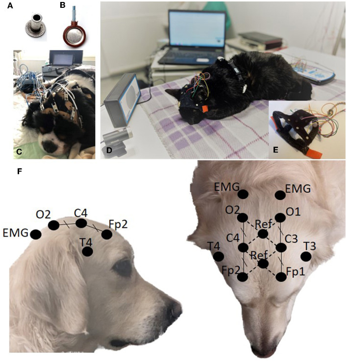
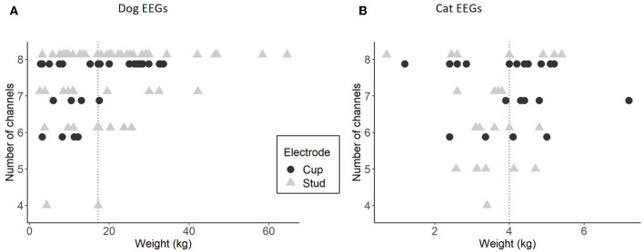
Procedures: We compared recordings performed with 8 EEG unwired stud Ag/AgCl electrodes held by elastic straps and 8 EEG wired cup Ag electrodes held by a tailor-made manufactured headset combined with a wired video-EEG device. Electrodes placement was determined according to previously published animal EEG protocols. Physiological sensors, such as electrocardiography, electromyography, and respiratory sensors were added. Stimulation protocols were tested. Quality and interpretability were evaluated.

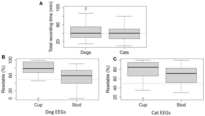
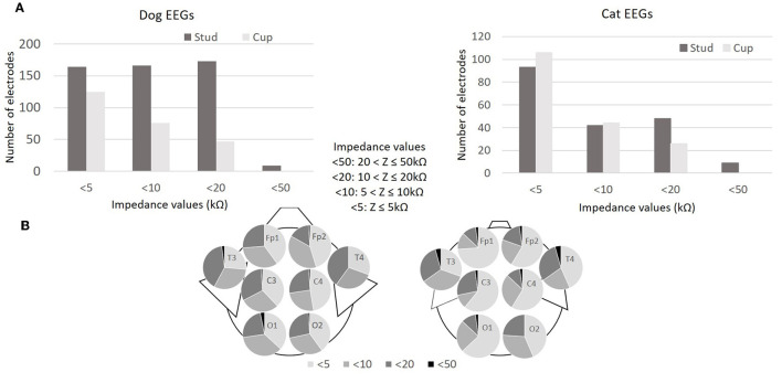
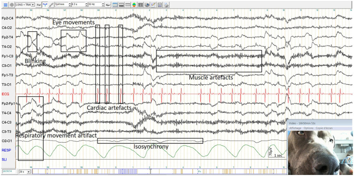
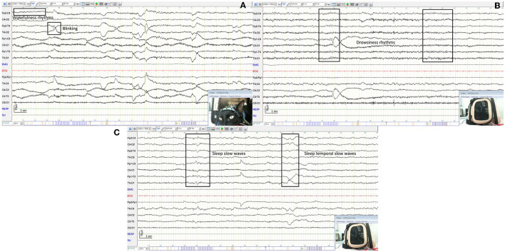
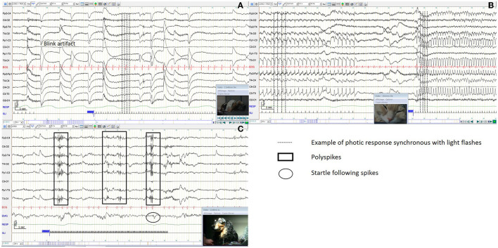
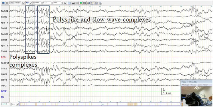
Results: Headsets and recording procedures appeared suitable for all skull shapes and sizes. Video-EEG recordings were successfully performed without tranquilization or anesthesia except for 9 animals. Median EEG recordings time was 40 min. Impedance remained below 20 kΩ in 99% of dog EEGs and 98% of cat EEGs. Isosynchrony was reported in 6% of the channels. Seventy-five percent of dog EEGs and 83% of cat EEGs were readable for more than 50% (to 100%) of their duration. Successful discrimination of vigilance states from rhythm analysis (wakefulness, drowsiness, and sleepiness) was possible in 99% of dog EEGs and 91% of cat EEGs. Photic driving responses during photic stimulations were observed in 11% of dog EEGs and 85% of cat EEGs. Electroencephalography recordings were directly informative in 32% of the examinations: in 25% EEG abnormalities were associated with clinical signs and 7% concerned EEG abnormalities without clinical symptoms during recording. Thirteen percent of dogs subjected to photic stimulation exhibited epileptic anomalies. Among 9 EEGs with other history-based stimulations, three displayed epileptic graphoelements.

Conclusions: We have developed a standardized unanesthetized video-EEG procedure easily performed and reproducible in dogs and cats. Qualitative and quantitative technical and medical criteria were evaluated and were in accordance with human EEG recommendations. Moreover, we have demonstrated its relevance and accuracy for diagnostic purposes, providing further arguments for the use of EEG as a first-line neurological functional exploration test.

Keywords: EEG; canine; encephalopathy; epilepsy; feline; gel electrode; video-EEG.


Conflict of interest statement

EL was employed by AMSET MEDICAL. EL is the patent applicant No. FR3101533. The remaining authors declare that the research was conducted in the absence of any commercial or financial relationships that could be construed as a potential conflict of interest.

Figures

Figure 1
Figure 1
Stud electrode (A). Cup electrode (B). EEG acquisition setup using stud electrodes and elastic straps (C). EEG acquisition setup using cup electrodes and PetCap® (D) with details of electrodes coated with conductive paste and inserted into the system (E). Electrode positioning (F).
Figure 2
Figure 2
Total recording time for dog EEGs (95) and cat EEGs (46) (A). Readable percentage calculated with the 20-second pages that are undisturbed by artifacts for more than half of their duration over the total recording time depending on electrode type, for dog EEGs (64 EEGs with stud electrodes and 31 EEGs with cup electrodes) (B) and cat EEGs (24 EEGs with stud electrodes and 22 EEGs with cup electrodes) (C).
Figure 3
Figure 3
Impedance values (kΩ) of the 760 electrodes during the 95 dog EEGs (95x8 electrodes) and impedance values of the 368 electrodes during the 46 cat EEGs (46x8 electrodes) (A). Distribution of impedance values (kΩ) according to electrode position in 95 dog EEGs and 46 cat EEGs (B).
Figure 4
Figure 4
Examples of isosynchrony and physiological artifacts in a dog EEG.
Figure 5
Figure 5
Relationship between the weight of the animals and the number of usable channels in longitudinal montage, for dog EEGs with stud (64 EEGs) and cup (31 EEGs) (A) and for cat EEGs with stud (24 EEGs) and cup (22 EEGs) (B). The dotted line represents the median of the weights in each species.
Figure 6
Figure 6
EEG of an awake dog showing a rapid low amplitude rhythm and artifacts from blinking and muscle activity with examples in boxes (A). EEG of the same dog as before, awake and calm, showing a rapid low amplitude rhythm with reduced artifacts from blinking and muscle activity with examples in boxes (B). EEG of the same dog as before, dozy, showing a 5 Hz medium amplitude rhythm (C). EEG of the same dog as before, sleepy, showing 1−3 Hz high amplitude rhythms with examples in boxes (D).
Figure 7
Figure 7
EEG of an awake cat showing a rapid low amplitude rhythm and artifacts from blinking and muscle activity with examples in boxes (A). EEG of the same cat as before, dozy, showing a 7 Hz medium amplitude rhythm (B). EEG of the same cat as before, sleepy, showing mixed theta and delta rhythms with examples in boxes (C).
Figure 8
Figure 8
Rhythm distinctions (W, wakefulness only; WD, wakefulness and drowsiness; WDS, wakefulness, drowsiness and sleeping; D, drowsiness only; O, other than physiological states) in the recordings of 95 dogs (on the left) and 46 cats (on the right) detailed according to the type of electrodes used and expressed as a percentage of the total number of recordings made for each kind of electrode, cup and stud (A), their sex (B), their age (C), and the total recording time (D).
Figure 9
Figure 9
Reactivity to intermittent photic stimulation in a dog EEG (A), in a cat EEG (B), with polyspike complexes in a dog EEG (C).
Figure 10
Figure 10
Polyspike complexes and polyspike-and-slow-wave-complexes and simultaneous twitching of a cat's face.

References

    1. Scheffer IE, Berkovic S, Capovilla G, Connolly MB, French J, Guilhoto L, et al. . ILAE classification of the epilepsies: position paper of the ILAE Commission for Classification and Terminology. Epilepsia. (2017) 58:512–21. 10.1111/epi.13709 - DOI - PMC - PubMed
    1. Nuwer MR, Comi G, Emerson R, Fuglsang-Frederiksen A, Guérit JM, Hinrichs H, et al. . IFCN standards for digital recording of clinical EEG. Electroencephalogr Clin Neurophysiol. (1998) 106:259–61. 10.1016/S0013-4694(97)00106-5 - DOI - PubMed
    1. Flink R, Pedersen B, Guekht AB, Malmgren K, Michelucci R, Neville B, et al. . Guidelines for the use of EEG methodology in the diagnosis of epilepsy. Acta Neurol Scand. (2002) 106:1–7. 10.1034/j.1600-0404.2002.01361.x - DOI - PubMed
    1. American Clinical Neurophysiology Society . Guideline 2: Minimum technical standards for pediatric electoencephalography. J Clin Neurophysiol Off Publ Am Electroencephalogr Soc. (2006) 23:92–6. 10.1097/00004691-200604000-00003 - DOI - PubMed
    1. André-Obadia N, Sauleau P, Cheliout-Heraut F, Convers P, Debs R, Eisermann M, et al. . Recommandations françaises sur l'électroencéphalogramme. Neurophysiol Clin Neurophysiol. (2014) 44:515–612. 10.1016/j.neucli.2014.10.001 - DOI - PubMed