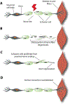
Biomimetic Strategies for Peripheral Nerve Injury Repair: An Exploration of Microarchitecture and Cellularization
- PMID: 38343513
- PMCID: PMC10857769
- DOI: 10.1007/s44174-022-00039-8
Biomimetic Strategies for Peripheral Nerve Injury Repair: An Exploration of Microarchitecture and Cellularization
Abstract
Injuries to the nervous system present formidable challenges to scientists, clinicians, and patients. While regeneration within the central nervous system is minimal, peripheral nerves can regenerate, albeit with limitations. The regenerative mechanisms of the peripheral nervous system thus provide fertile ground for clinical and scientific advancement, and opportunities to learn fundamental lessons regarding nerve behavior in the context of regeneration, particularly the relationship of axons to their support cells and the extracellular matrix environment. However, few current interventions adequately address peripheral nerve injuries. This article aims to elucidate areas in which progress might be made toward developing better interventions, particularly using synthetic nerve grafts. The article first provides a thorough review of peripheral nerve anatomy, physiology, and the regenerative mechanisms that occur in response to injury. This is followed by a discussion of currently available interventions for peripheral nerve injuries. Promising biomaterial fabrication techniques which aim to recapitulate nerve architecture, along with approaches to enhancing these biomaterial scaffolds with growth factors and cellular components, are then described. The final section elucidates specific considerations when developing nerve grafts, including utilizing induced pluripotent stem cells, Schwann cells, nerve growth factors, and multilayered structures that mimic the architectures of the natural nerve.
Keywords: Cellularized pathways; Nerve guidance conduit; Peripheral nerve regeneration; Synthetic scaffolds.
Conflict of interest statement
Conflict of interest The authors have no competing interests to declare that are relevant to the content of this article.
Figures


Similar articles
-
Advances in Biomimetic Nerve Guidance Conduits for Peripheral Nerve Regeneration.Nanomaterials (Basel). 2023 Sep 10;13(18):2528. doi: 10.3390/nano13182528. Nanomaterials (Basel). 2023. PMID: 37764557 Free PMC article. Review.
-
Advances and clinical challenges for translating nerve conduit technology from bench to bed side for peripheral nerve repair.Cell Tissue Res. 2021 Feb;383(2):617-644. doi: 10.1007/s00441-020-03301-x. Epub 2020 Nov 17. Cell Tissue Res. 2021. PMID: 33201351 Review.
-
Engineering a multimodal nerve conduit for repair of injured peripheral nerve.J Neural Eng. 2013 Feb;10(1):016008. doi: 10.1088/1741-2560/10/1/016008. Epub 2013 Jan 3. J Neural Eng. 2013. PMID: 23283383
-
Construction of tissue engineered nerve grafts and their application in peripheral nerve regeneration.Prog Neurobiol. 2011 Feb;93(2):204-30. doi: 10.1016/j.pneurobio.2010.11.002. Epub 2010 Dec 2. Prog Neurobiol. 2011. PMID: 21130136 Review.
-
Biomimetic Architectures for Peripheral Nerve Repair: A Review of Biofabrication Strategies.Adv Healthc Mater. 2018 Apr;7(8):e1701164. doi: 10.1002/adhm.201701164. Epub 2018 Jan 19. Adv Healthc Mater. 2018. PMID: 29349931 Review.
Cited by
-
Motor Function in the Setting of Nerve Allografts: Is This the Future of Facial Nerve Reconstruction?J Clin Med. 2025 Aug 5;14(15):5510. doi: 10.3390/jcm14155510. J Clin Med. 2025. PMID: 40807130 Free PMC article. Review.
-
Biomaterials in tissue repair and regeneration: key insights from extracellular matrix biology.Front Med Technol. 2025 Aug 15;7:1565810. doi: 10.3389/fmedt.2025.1565810. eCollection 2025. Front Med Technol. 2025. PMID: 40893932 Free PMC article. Review.
References
-
- Houdek MT, Shin AY, Management and complications of traumatic peripheral nerve injuries. Hand Clin. 31(2), 151–163 (2015) - PubMed
-
- Brattain K, analysis of the peripheral nerve repair market in the United States, (2013).
Grants and funding
LinkOut - more resources
Full Text Sources
