. 2024 Jan 19;20(3):42.
doi: 10.3892/br.2024.1727. eCollection 2024 Mar.

Irreversible inhibition of estrogen receptor α signaling and the emergence of hormonal resistance in MCF7 breast cancer cells induced by DNA damage agents

Affiliations

Irreversible inhibition of estrogen receptor α signaling and the emergence of hormonal resistance in MCF7 breast cancer cells induced by DNA damage agents

Alexander M Scherbakov et al. Biomed Rep. .

Abstract

Combining chemotherapy and hormone therapy is a prevalent approach in breast cancer treatment. While the cytotoxic impact of numerous chemotherapy drugs stems from DNA damage, the exact role of these DNA alterations in modulating estrogen receptor α (ERα) machinery remains elusive. The present study aimed to analyze the impact of DNA damage agents on ERα signaling in breast cancer cells and assess the signaling pathways mediating the influence of DNA damage drugs on the ERα machinery. Cell viability was assessed using the MTT method, while the expression of signaling proteins was analyzed by immunoblotting. ERα activity in the cells treated with various drugs (17β-estradiol, tamoxifen, 5-fluorouracil) was assessed through reporter gene assays. In vitro experiments were conducted on MCF7 breast cancer cells subjected to varying durations of 5-fluorouracil (5-FU) treatment. Two distinct cell responses to 5-FU were identified based on the duration of the treatment. A singular dose of 5-FU induces pronounced DNA fragmentation, temporally suppressing ERα signaling while concurrently activating AKT phosphorylation. This suppression reverses upon 5-FU withdrawal, restoring normalcy within ten days. However, chronic 5-FU treatment led to the emergence of 5-FU-resistant cells with irreversible alterations in ERα signaling, resulting in partial hormonal resistance. These changes mirror those observed in cells subjected to UV-induced DNA damage, underscoring the pivotal role of DNA damage in shaping estrogen signaling alterations in breast cancer cells. In summary, the results of the present study suggested that the administration of DNA damage agents to cancer cells can trigger irreversible suppression of estrogen signaling, fostering the development of partial hormonal resistance. This outcome may ultimately impede the efficacy of combined or subsequent chemo- and hormone therapy strategies.

Keywords: 5-fluorouracil; DNA damage; breast cancer; estrogen receptor α; progression; resistance; tamoxifen; ultraviolet C irradiation.


Conflict of interest statement

The authors declare that they have no competing interests.

Figures

Figure 1
Figure 1
MCF7 cells response to 5-FU. (A) The MCF7 cells were treated with 5-FU at the indicated doses and the efficiency of 5-FU-induced DNA damage was evaluated using the DNA fragmentation test-Comet assay, and by the accumulation of micronuclei in cells. The significance of differences in the damage level between untreated and treated cells was calculated using Pearson's χ2 test. (B) The sensitivity of MCF7 cells to 5-FU treatment. MCF7 cells were treated with 1.25-20 µM 5-FU for three days and the cell viability was assessed by the MTT assay. Data represent the mean value ± SD of three independent experiments. Percentage of 100% was set as the viability of MCF7 cells treated with vehicle control. **P<0.01 and ***P<0.0001. 5-FU, 5-fluorouracil.
Figure 2
Figure 2
5-FU treatment and ERα signaling in MCF7 cells. (A) Western blot analysis of ERα, p-AKT, AKT, p-p85/p-p70 S6K and p85/p70 S6K in cell extracts. The MCF7 cells were treated with 15 µM 5-FU for three days and the cells were subjected to western blotting. Protein loading was controlled by membrane hybridization with α-tubulin antibodies. The blot represents the results of one of three similar experiments. Densitometry for the tested proteins/α-tubulin ratio was carried out using ImageJ software (right diagram). *P<0.05. (B) Reporter analysis of ERα transcriptional activity. The cells were treated with 15 µM 5-FU for three days, then the cells were transfected with the plasmid containing the luciferase reporter gene under estrogen-responsive elements, and β-galactosidase plasmid. The cells were treated with or without 10 nM 17β-estradiol (E2) for 24 h, and the luciferase and β-galactosidase activities were determined. The relative luciferase activity was calculated in arbitrary units as the ratio of luciferase to the β-galactosidase activity. A total of 100 relative units were set as the luciferase activity in MCF7 cells treated with E2. Data represent the mean values ± S.D. of three independent experiments: *P<0.05 vs. untreated samples; #P<0.05 vs. E2-treated MCF7 cells. (C) Cell sensitivity to antiestrogen tamoxifen. The cells were treated with 15 µM 5-FU for three days following 5-FU withdrawal for three days. Then MCF7 cells were treated with 5 µM tamoxifen for three days and the number of viable cells was assessed by the MTT-test. Data represent the mean value ± SD of three independent experiments. Percentage of 100% was set as the viability of untreated cells. *P<0.05 vs. untreated samples. (D) Analysis of luciferase activity in MCF7 cells after 5-FU withdrawal. The cells were treated with 15 µM 5-FU for three days following 5-FU withdrawal for ten days: *P<0.05 vs. untreated samples. (E) The cell response to tamoxifen. *P<0.05 vs. untreated samples. (F) Western blot analysis of ERα, p-AKT, AKT, p-S6K, and S6K in control MCF7 cells and MCF7 cells 10 days after treatment. 5-FU, 5-fluorouracil; ERα, estrogen receptor α; 5-FU, 5-fluorouracil; S6K, Ribosomal Protein S6 Kinase B1; p-, phosphorylated.
Figure 3
Figure 3
Prolonged 5-FU treatment and selection of 5-FU-resistant cells. The MCF7 cells were treated with 15 µM 5-FU within two months with subsequent 5-FU withdrawal and cell cultivation in medium without drug for the next one month. (A and B) The sensitivity of the established MCF7/5-FUR cells to (A) 5-FU, (B) tamoxifen and (C) DNA damage tests (Comet assay and accumulation of micronuclei). (D and E) ERα signaling in 5-FU-resistant MCF7/5-FUR cells. (D) Reporter analysis of ERα and (E) western blot analysis of ERα, p-AKT, AKT, p-S6K, and S6K expression in MCF7 and MCF7/5-FUR cells. *P<0.05 and **P<0.01 vs. untreated samples; #P<0.05 vs. E2-treated MCF7 cells activity. ERα, estrogen receptor α; 5-FU, 5-fluorouracil; 5-FUR, 5-FU resistant; S6K, Ribosomal Protein S6 Kinase B1; TAM, tamoxifen; p-, phosphorylated.
Figure 4
Figure 4
UV influence on the viability of MCF7 cells. (A and B) The cells were exposed to a single UVC dose, and after 0-24 h (A) Comet assay (the significance of differences in the damage level between control and UV-exposed cells was calculated using Pearson's χ2 test; *P<0.05 vs. untreated and ‘0 h’ samples) and (B) colony-forming test (*P<0.05 vs. control samples) (b) were performed. UV, ultraviolet.
Figure 5
Figure 5
UVC influence on ERα signaling in MCF7 cells. (A-C) The cells were exposed to a single UVC dose; (A) western blot analysis of ERα, p-AKT, AKT, p-S6K and S6K (1 day after treatment, *P<0.05 vs. MCF7/untreated), (B) reporter analysis of ERα (*P<0.05 vs. untreated samples; #P<0.05 vs. E2-treated MCF7 cells) and (C) cell response to tamoxifen (3 days treatment with tamoxifen) were performed. UV, ultraviolet; ERα, estrogen receptor α; S6K, Ribosomal Protein S6 Kinase B; E2, 17β-estradiol; TAM, tamoxifen; p-, phosphorylated.
Figure 6
Figure 6
Analysis of ERα signaling in MCF7 cells 30 days after single UVС irradiation. (A) Western blot analysis, (B) reporter analysis of ERα and (C) cell response to tamoxifen were performed; *P<0.05 vs. untreated samples. ERα, estrogen receptor α; UV, ultraviolet; S6K, Ribosomal Protein S6 Kinase B1; E2, 17β-estradiol; p-, phosphorylated.
Figure 7
Figure 7
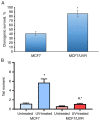
Selection and cell viability of UV-resistant subline. The MCF7 cells were exposed to UVC once every three days for 4 weeks with subsequent cell growth in standard medium for the next 40 days. (A and B) The comparative analysis of the cell viability of the parent MCF7 cells and the established MCF7/UVR subline was performed using (A) colony-forming test (*P<0.05 vs. MCF7 cells) and (B) Comet assay (the difference between control and UV-exposed cells was calculated using Pearson's χ2 test) (*P<0.05 vs. untreated cells; #P<0.05 vs. UV-treated MCF7 cells). UV, ultraviolet.
Figure 8
Figure 8
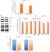
ERα signaling in MCF7/UVR cells. (A) Reporter analysis of ERα (*P<0.05 vs. untreated samples; #P<0.05 vs. E2-treated MCF7 cells), (B) western blot analysis and (C) cell sensitivity to tamoxifen (*P<0.05 vs. untreated samples). ERα, estrogen receptor α; UV, ultraviolet; p-, phosphorylated.

Similar articles

References

    1. Tiezzi DG, De Andrade JM, Cândido dos Reis FJ, Marana HR, Ribeiro-Silva A, Tiezzi MG, Pereira AP. Apoptosis induced by neoadjuvant chemotherapy in breast cancer. Pathology. 2006;38:21–27. doi: 10.1080/00313020500465315. - DOI - PubMed
    1. Khan M, Rasul A, Yi F, Zhong L, Ma T. Jaceosidin induces p53-dependent G2/M phase arrest in U87 glioblastoma cells. Asian Pac J Cancer Prev. 2011;12:3235–3238. - PubMed
    1. Rasul A, Khan M, Yu B, Ma T, Yang H. Xanthoxyletin, a coumarin induces S phase arrest and apoptosis in human gastric adenocarcinoma SGC-7901 cells. Asian Pac J Cancer Prev. 2011;12:1219–1223. - PubMed
    1. Ding M, Shao Y, Sun D, Meng S, Zang Y, Zhou Y, Li J, Lu W, Zhu S. Design, synthesis, and biological evaluation of BRD4 degraders. Bioorg Med Chem. 2023;78(117134) doi: 10.1016/j.bmc.2022.117134. - DOI - PubMed
    1. Chrienova Z, Rysanek D, Oleksak P, Stary D, Bajda M, Reinis M, Mikyskova R, Novotny O, Andrys R, Skarka A, et al. Discovery of small molecule mechanistic target of rapamycin inhibitors as anti-aging and anti-cancer therapeutics. Front Aging Neurosci. 2022;14(1048260) doi: 10.3389/fnagi.2022.1048260. - DOI - PMC - PubMed