

Immune checkpoint blockade improves the activation and function of circulating mucosal-associated invariant T (MAIT) cells in patients with non-small cell lung cancer
- PMID: 38343750
- PMCID: PMC10854269
- DOI: 10.1080/2162402X.2024.2312631
Immune checkpoint blockade improves the activation and function of circulating mucosal-associated invariant T (MAIT) cells in patients with non-small cell lung cancer
Abstract
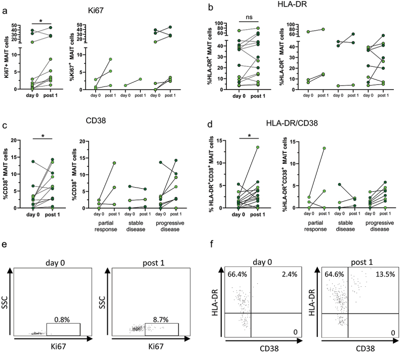
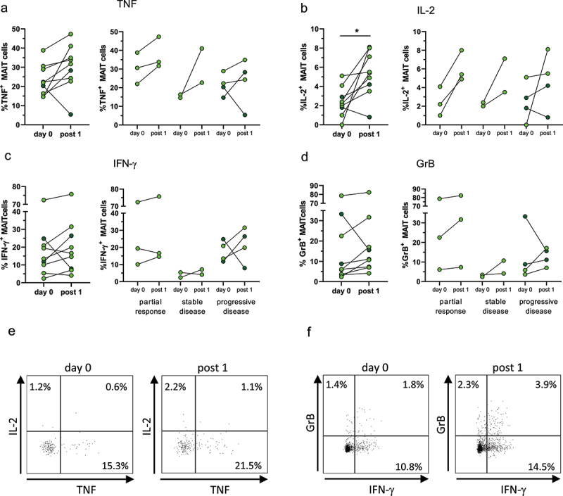
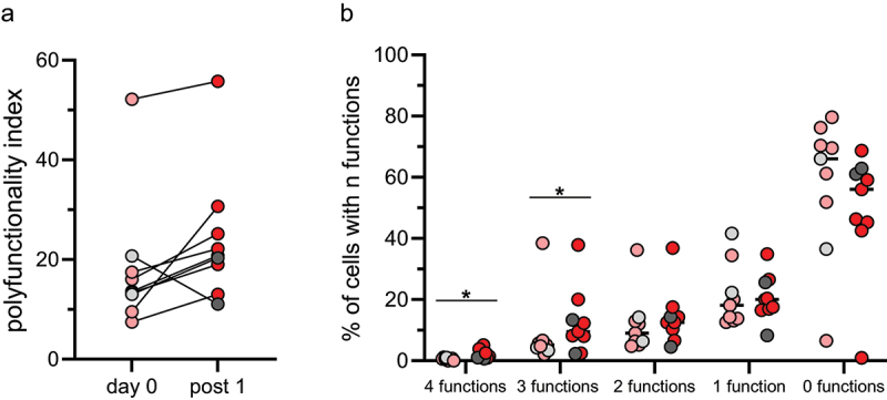
Mucosal-associated invariant T (MAIT) cells constitute one of the most numerous unconventional T cell subsets, and are characterized by rapid release of Th1- and Th17-associated cytokines and increased cytotoxic functions following activation. MAIT cells accumulate in tumor tissue but show an exhausted phenotype. Here, we investigated if immune checkpoint blockade (ICB) with antibodies to PD-1 or PD-L1 affects the function of circulating MAIT cells from non-small cell lung cancer patients. ICB increased the proliferation and co-expression of the activation markers HLA-DR and CD38 on MAIT cells in most patients after the first treatment cycle, irrespective of treatment outcome. Furthermore, production of cytokines, especially TNF and IL-2, also increased after treatment, as did MAIT cell polyfunctionality. These results indicate that MAIT cells respond to ICB, and that MAIT cell reinvigoration may contribute to tumor regression in patients undergoing immune checkpoint therapy.
Keywords: Immune checkpoint blockade; MAIT cell; PD-1; immunotherapy; non-small cell lung cancer.
© 2024 The Author(s). Published with license by Taylor & Francis Group, LLC.
Conflict of interest statement
M Quiding Järbrink has received consultancy fees from Biomunex Pharmaceuticals.
Figures
Similar articles
-
Mucosal-associated invariant T-cells in pulmonary pathophysiology.Curr Opin Pulm Med. 2025 May 1;31(3):202-210. doi: 10.1097/MCP.0000000000001163. Epub 2025 Mar 19. Curr Opin Pulm Med. 2025. PMID: 40104908 Free PMC article. Review.
-
CXCR6-positive circulating mucosal-associated invariant T cells can identify patients with non-small cell lung cancer responding to anti-PD-1 immunotherapy.J Exp Clin Cancer Res. 2024 May 3;43(1):134. doi: 10.1186/s13046-024-03046-3. J Exp Clin Cancer Res. 2024. PMID: 38698468 Free PMC article.
-
Clinicopathological and predictive value of MAIT cells in non-small cell lung cancer for immunotherapy.J Immunother Cancer. 2023 Jan;11(1):e005902. doi: 10.1136/jitc-2022-005902. J Immunother Cancer. 2023. PMID: 36657812 Free PMC article.
-
Exhaustion in tumor-infiltrating Mucosal-Associated Invariant T (MAIT) cells from colon cancer patients.Cancer Immunol Immunother. 2021 Dec;70(12):3461-3475. doi: 10.1007/s00262-021-02939-y. Epub 2021 Apr 22. Cancer Immunol Immunother. 2021. PMID: 33885944 Free PMC article.
-
Mucosal-Associated Invariant T cell in liver diseases.Int J Biol Sci. 2020 Jan 1;16(3):460-470. doi: 10.7150/ijbs.39016. eCollection 2020. Int J Biol Sci. 2020. PMID: 32015682 Free PMC article. Review.
Cited by
-
Mucosal-associated invariant T-cells in pulmonary pathophysiology.Curr Opin Pulm Med. 2025 May 1;31(3):202-210. doi: 10.1097/MCP.0000000000001163. Epub 2025 Mar 19. Curr Opin Pulm Med. 2025. PMID: 40104908 Free PMC article. Review.
-
Biological and clinical significance of circulating mucosal-associated invariant T cells in lung cancer.Transl Cancer Res. 2025 Mar 30;14(3):1995-2009. doi: 10.21037/tcr-2025-178. Epub 2025 Mar 27. Transl Cancer Res. 2025. PMID: 40224974 Free PMC article.
-
Unconventional T cells in anti-cancer immunity.Front Immunol. 2025 Jul 17;16:1618393. doi: 10.3389/fimmu.2025.1618393. eCollection 2025. Front Immunol. 2025. PMID: 40746558 Free PMC article. Review.
References
-
- Yan J, Allen S, McDonald E, Das I, Mak JYW, Liu L, Fairlie DP, Meehan BS, Chen Z, Corbett AJ, et al. MAIT cells promote tumor initiation, growth, and metastases via tumor MR1. Cancer Discov. 2020;10(1):124–141. doi: 10.1158/2159-8290.CD-19-0569. - DOI - PubMed
MeSH terms
Substances
LinkOut - more resources
Full Text Sources
Medical
Research Materials