Review
. 2024 Feb 9:15:20417314241228118.
doi: 10.1177/20417314241228118. eCollection 2024 Jan-Dec.

Biosubstitutes for dural closure: Unveiling research, application, and future prospects of dura mater alternatives

Affiliations
Review

Biosubstitutes for dural closure: Unveiling research, application, and future prospects of dura mater alternatives

Dolphee Khurana et al. J Tissue Eng. .

Abstract

The dura mater, as the crucial outermost protective layer of the meninges, plays a vital role in safeguarding the underlying brain tissue. Neurosurgeons face significant challenges in dealing with trauma or large defects in the dura mater, as they must address the potential complications, such as wound infections, pseudomeningocele formation, cerebrospinal fluid leakage, and cerebral herniation. Therefore, the development of dural substitutes for repairing or reconstructing the damaged dura mater holds clinical significance. In this review we highlight the progress in the development of dural substitutes, encompassing autologous, allogeneic, and xenogeneic replacements, as well as the polymeric-based dural substitutes fabricated through various scaffolding techniques. In particular, we explore the development of composite materials that exhibit improved physical and biological properties for advanced dural substitutes. Furthermore, we address the challenges and prospects associated with developing clinically relevant alternatives to the dura mater.

Keywords: Dura mater; brain trauma and injury; composites; dural substitutes; polymeric scaffolds; tissue engineering.


Conflict of interest statement

The author(s) declared no potential conflicts of interest with respect to the research, authorship, and/or publication of this article.

Figures

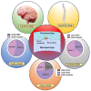
Graphical abstract
Graphical abstract
Figure 1.
Figure 1.
Illustrates a schematic representation of the dural substitutes currently employed for dural repair.
Figure 2.
Figure 2.
Schematic representation showing the dural defect and the available substitutes for dura mater repair. It also presents the relative percentage of studies conducted on dural grafts categorized by year.,,,,, –
Figure 3.
Figure 3.
Detailed representation of the dura mater present in the cranium and spine: (a) a diagram showing the meninges of the human head which includes the dura mater, the arachnoid mater and the pia, (b) a close-up view of the cranial dura mater. Copyright 2021, Scientific Research Publishing, (c) a diagram of the spinal cord showing the dura mater as a protective covering. Copyright 2022, American Society of Neuroradiology. (d) A close-up view of the cranial dura mater. Copyright 2021, Scientific Research Publishing.
Figure 4.
Figure 4.
A schematic representation of the cranial meninges and transverse section of the dura mater as seen in the SEM image where (a) is the bone surface layer, (b) represents the external median layer, (c) is the vascular layer, (d) is internal median layer, and (e) represents the arachnoid layer.
Figure 5.
Figure 5.
A schematic overview of the preparation, implantation, and regeneration process of the dehydarated amniotic membrane: illustrative depiction of the amniotic membrane as a viable substitute for autografts in the context of dura mater reconstruction or repair.
Figure 6.
Figure 6.
Demonstration of the ZYMO-TECK® involved in the processing of Heart patch and the surgical process involved in the implantation: (a) illustrative diagram showcasing the equine pericardium membrane as a xenogeneic solution for replacing the dura mater. Heart® membrane employed as a dural alternative, (b) the membrane positioned over the defect, (c) the membrane cut into appropriate dimensions, and (d) the membrane sutured to the patient’s dura mater. Copyright 2016, Thieme Medical Publishers.
Figure 7.
Figure 7.
Illustrative depiction of the origin and morphology of pericardial patch and Biodesign® along with the duraplasty procedure: (a) schematic representation of pericardial patch and Biodesign® dural repair graft-based approach for duraplasty, (b) wet form of the porcine pericardial patch, (c) dry form of the Biodesign® Dural Repair Graft, (d) performance of bilateral craniotomies, and (e) duraplasty done using each type of dural substitute. Copyright 2018, Society of Neuroscience.
Figure 8.
Figure 8.
The production method for a dual-layer biomaterial composed of silk fibroin involves the following steps.
Figure 9.
Figure 9.
Schematic diagram of the fabrication of triple-layered PCL dura mater substitute (loaded with GS and nHA): (a) electrohydrodynamic fabrication of PCL-GS-nHA dura mater scaffold, (b) electrospinning of highly aligned PCL-GS nanofibers, (c) electrospinning of random PCL-GS nanofibers, and (d) fabrication of melt-based microscale PCL-nHA fibers. Copyright 2022, Elsevier.
Figure 10.
Figure 10.
Systematic images show that the triple-layered composite has effective multifunctionality, including leak-proof ability, antiadhesion performance, antibacterial ability, and dura regeneration potential. Copyright 2021, Elsevier.
Figure 11.
Figure 11.
Graphical representation of the mechanical properties and proportion of studies performed on various types of grafts: (a) ultimate tensile strength for various dural substitutes,,,,,,,,,, (b) Young’s modulus for different dural substitutes,,,,, and (c) ratio clinical, in vivo and in vitro study for the dural grafts.,,,,, –,

Similar articles

Cited by

References

    1. Yu X, Yue P, Peng X, et al.. A dural substitute based on oxidized quaternized guar gum/porcine peritoneal acellular matrix with improved stability, antibacterial and anti-adhesive properties. Chin Chem Lett 2023; 34: 107591.
    1. Chuan D, Wang Y, Fan R, et al.. Fabrication and properties of a biomimetic dura matter substitute based on stereocomplex poly (Lactic acid) nanofibers. Int J Nanomedicine 2020; 15: 3729–3740. - PMC - PubMed
    1. Song Y, Li S, Song B, et al.. The pathological changes in the spinal cord after dural tear with and without autologous fascia repair. Eu Spine J 2014; 23: 1531–1540. - PubMed
    1. Wang W, Ao Q. Research and application progress on dural substitutes. J Neurorestoratol 2019; 7: 161–170.
    1. Jackson N, Muthuswamy J. Artificial dural sealant that allows multiple penetrations of implantable brain probes. J Neurosci Methods 2008; 171: 147–152. - PMC - PubMed

LinkOut - more resources