

This is a preprint.
Advanced magnetic resonance imaging detects altered placental development in pregnancies affected by congenital heart disease
- PMID: 38343847
- PMCID: PMC10854304
- DOI: 10.21203/rs.3.rs-3873412/v1
Advanced magnetic resonance imaging detects altered placental development in pregnancies affected by congenital heart disease
Update in
-
Advanced magnetic resonance imaging detects altered placental development in pregnancies affected by congenital heart disease.Sci Rep. 2024 May 29;14(1):12357. doi: 10.1038/s41598-024-63087-8. Sci Rep. 2024. PMID: 38811636 Free PMC article.
Abstract
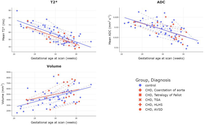
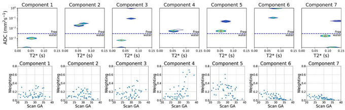
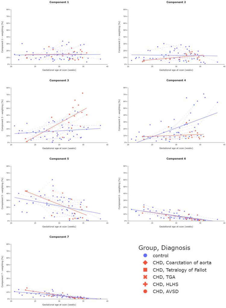
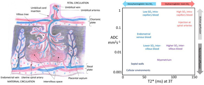
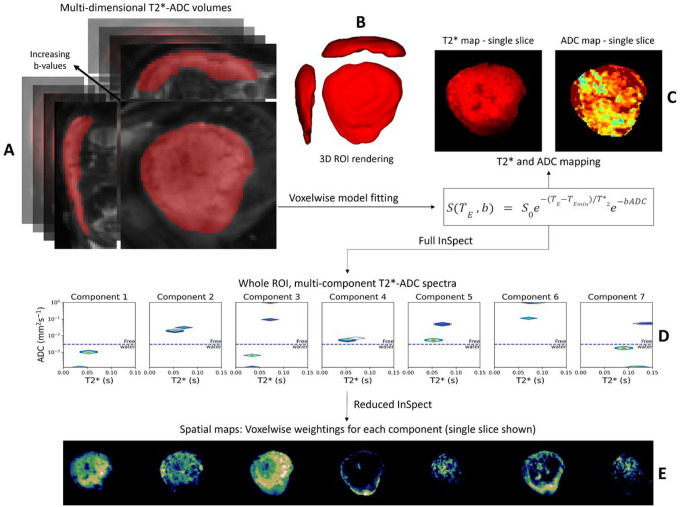
Congenital heart disease (CHD) is the most common congenital malformation and is associated with adverse neurodevelopmental outcomes. The placenta is crucial for healthy fetal development and placental development is altered in pregnancy when the fetus has CHD. This study utilized advanced combined diffusion-relaxation MRI and a data-driven analysis technique to test the hypothesis that placental microstructure and perfusion are altered in CHD-affected pregnancies. 48 participants (36 controls, 12 CHD) underwent 67 MRI scans (50 control, 17 CHD). Significant differences in the weighting of two independent placental and uterine-wall tissue components were identified between the CHD and control groups (both pFDR<0.001), with changes most evident after 30 weeks gestation. A Significant trend over gestation in weighting for a third independent tissue component was also observed in the CHD cohort (R = 0.50, pFDR=0.04), but not in controls. These findings add to existing evidence that placental development is altered in CHD. The results may reflect alterations in placental perfusion or the changes in fetal-placental flow, villous structure and maturation that occur in CHD. Further research is needed to validate and better understand these findings and to understand the relationship between placental development, CHD, and its neurodevelopmental implications.
Conflict of interest statement
Competing interests: The author(s) declare no competing interests.
Figures
Similar articles
-
Advanced magnetic resonance imaging detects altered placental development in pregnancies affected by congenital heart disease.Sci Rep. 2024 May 29;14(1):12357. doi: 10.1038/s41598-024-63087-8. Sci Rep. 2024. PMID: 38811636 Free PMC article.
-
3-D volumetric MRI evaluation of the placenta in fetuses with complex congenital heart disease.Placenta. 2015 Sep;36(9):1024-30. doi: 10.1016/j.placenta.2015.06.013. Epub 2015 Jul 6. Placenta. 2015. PMID: 26190037 Free PMC article.
-
Non-Invasive Placental Perfusion Imaging in Pregnancies Complicated by Fetal Heart Disease Using Velocity-Selective Arterial Spin Labeled MRI.Sci Rep. 2017 Nov 23;7(1):16126. doi: 10.1038/s41598-017-16461-8. Sci Rep. 2017. PMID: 29170468 Free PMC article.
-
Neuroplacentology in congenital heart disease: placental connections to neurodevelopmental outcomes.Pediatr Res. 2022 Mar;91(4):787-794. doi: 10.1038/s41390-021-01521-7. Epub 2021 Apr 16. Pediatr Res. 2022. PMID: 33864014 Free PMC article. Review.
-
Placental abnormalities in congenital heart disease.Transl Pediatr. 2021 Aug;10(8):2148-2156. doi: 10.21037/tp-20-347. Transl Pediatr. 2021. PMID: 34584887 Free PMC article. Review.
References
-
- Heazell A. The placenta and adverse pregnancy outcomes – opening the black box? BMC Pregnancy Childbirth 15(Suppl 1), (2015).
-
- Roseboom T. J. & Watson E. D. The next generation of disease risk: Are the effects of prenatal nutrition transmitted across generations? Evidence from animal and human studies. Placenta 33, e40–e44 (2012). - PubMed
-
- EUROCAT. European Platform on Rare Disease Registration. (2020).
-
- Latal B. Neurodevelopmental Outcomes of the Child with Congenital Heart Disease. Clin. Perinatol. 43, 173–185 (2016). - PubMed
Publication types
Grants and funding
LinkOut - more resources
Full Text Sources
