

Rare origin - Ewing's sarcoma of the pleura: a case report and literature review
- PMID: 38343883
- PMCID: PMC10852051
- DOI: 10.3941/jrcr.v17i8.5187
Rare origin - Ewing's sarcoma of the pleura: a case report and literature review
Abstract
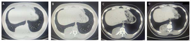
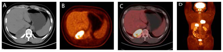
Ewing sarcoma (ES) was first reported by Ewing in 1921. It is the second largest malignant bone tumor in children and adolescents, typically occurring in the bones of trunk or limbs . Extraskeletal Ewing sarcoma (EES) was first reported by Tefft et al. in 1969 and is extremely rare, accounting for less than 1% of all sarcomas. It can occur in any part of soft tissue, mostly in the trunk and lower limbs, and rarely in the pleura. We report a 22-year-old case of extraosseous Ewing sarcoma of pleural origin discovered and pathologically confirmed by physical examination. We report its CT manifestations and pathological results, and review the literature to summarize and analyze the clinical and imaging characteristics of extraosseous Ewing sarcoma, in order to improve our understanding of the disease.
Copyright Journal of Radiology Case Reports.
Figures
Similar articles
-
Esophageal extraskeletal neoplasm Ewing's sarcoma: Case report.Int J Surg Case Rep. 2022 Aug;97:107399. doi: 10.1016/j.ijscr.2022.107399. Epub 2022 Jul 9. Int J Surg Case Rep. 2022. PMID: 35926382 Free PMC article.
-
Extraosseous Ewing's sarcoma.Can Assoc Radiol J. 1995 Apr;46(2):131-3. Can Assoc Radiol J. 1995. PMID: 7704678
-
A primary Ewing's sarcoma of pleura: Case report and literature review.Respir Med Case Rep. 2021 Sep 13;34:101516. doi: 10.1016/j.rmcr.2021.101516. eCollection 2021. Respir Med Case Rep. 2021. PMID: 34584837 Free PMC article.
-
[Ewing sarcoma. Diagnostic imaging].Radiologe. 1998 Jun;38(6):509-22. doi: 10.1007/s001170050386. Radiologe. 1998. PMID: 9700772 Review. German.
-
Extraskeletal Ewing's sarcoma: a case report and review of the literature.Spine (Phila Pa 1976). 2000 Aug 1;25(15):1996-9. doi: 10.1097/00007632-200008010-00022. Spine (Phila Pa 1976). 2000. PMID: 10908947 Review.
Cited by
-
Primary intracranial extraosseous Ewing's sarcoma with intraspinal metastasis in children: illustrative case.J Neurosurg Case Lessons. 2025 Jan 6;9(1):CASE24488. doi: 10.3171/CASE24488. Print 2025 Jan 6. J Neurosurg Case Lessons. 2025. PMID: 39761549 Free PMC article.
References
Publication types
MeSH terms
LinkOut - more resources
Full Text Sources
Medical