Case Reports
. 2024 Jan 26:14:1194307.
doi: 10.3389/fcimb.2024.1194307. eCollection 2024.

Exploring the relationship of supernumerary recurrent renal calculi formation and tick-borne infections: a case report

Affiliations
Case Reports

Exploring the relationship of supernumerary recurrent renal calculi formation and tick-borne infections: a case report

Dean C Paz et al. Front Cell Infect Microbiol. .

Abstract

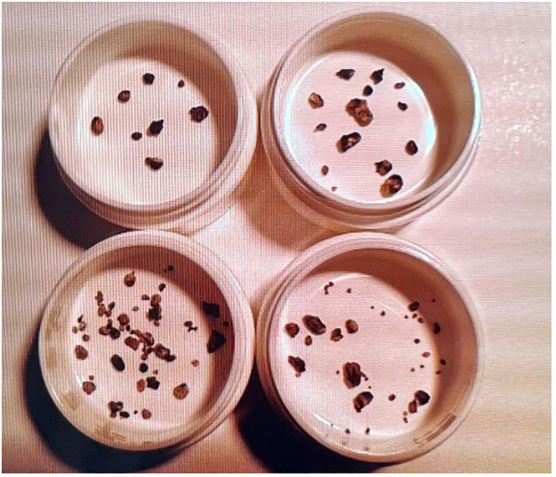
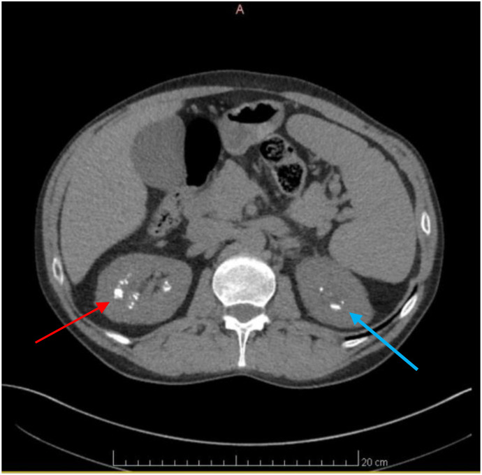
A 51-year-old male with a history of Cacchi-Ricci disease and long-standing infection with various species of Borrelia, Babesia, and Bartonella presented with recurrent symptoms of right-sided flank pain. Numerous renal calculi were identified on imaging. The etiology of the calculi had not been previously elucidated. Symptoms intermittently date back to 2002 when uric acid stones were identified. Subsequent calculi analysis revealed calcium oxalate stones. Despite the commonality of nephrolithiasis in patients with Cacchi-Ricci disease, the extreme number of calculi and recurrent presentation of symptoms persisted despite a plethora of medical evaluations, dietary changes, and hereditary testing. This case raises questions of etiology including possible immune deficiency and whether his uncommon microbial history contributes to recurrent stone formation.

Keywords: Babesia; Bartonella; Borrelia; Cacchi-Ricci disease; medullary sponge kidney; nephrolithiasis; renal calculi; tick-borne infections.


Conflict of interest statement

The authors declare that the research was conducted in the absence of any commercial or financial relationships that could be construed as a potential conflict of interest.

Figures

Figure 1
Figure 1
Renal calculi throughout the years. Microscopic image showcasing the assortment of renal calculi collected over several years. The top 2 containers are prior to 2021. The bottom left container is from 2021, and the bottom right corner is from 2022. The image highlights a multitude of supernumerary renal calculi, elucidating their recurrent formation. The correlation between these calculi and tick-borne infections is explored in this case report, presenting a potential link worthy of further investigation.
Figure 2
Figure 2
Line graph showing an increase in Vitamin D levels following supplementation. Graphical representation illustrating the patient’s dynamic Vitamin D levels over time. The patient initially had a level of 20ng/mL (normal range between 20 to 40ng/mL) on October 28th 2022, as indicated by the green arrow. The graph demonstrates a significant rise in Vitamin D levels following supplementation. From an initial deficient state, the patient’s Vitamin D levels increased to 33 ng/mL, as seen by the orange arrow, showcasing the impact of Vitamin D supplementation. This observation is pertinent to the investigation into the relationship between supernumerary recurrent renal calculi formation and potential associations with tick-borne infections, offering insights into the potential role of Vitamin D in this context.
Figure 3
Figure 3
Line graph showing a decrease in PTH following Vitamin D supplementation. Bar graph showing the impact of Vitamin D supplementation on Parathyroid Hormone (PTH) levels in our patient. The graph displays a noticeable decrease from an initial PTH level of 92 pg/mL, as seen by the grey arrow, to 83 pg/mL, as shown by the black arrow, following Vitamin D supplementation. This decline in PTH levels suggests a regulatory effect of Vitamin D on parathyroid function; Vitamin D plays a pivotal role in calcium homeostasis. When Vitamin D levels are low, there is a resultant rise in PTH secretion from the parathyroid glands, aiming to increase calcium absorption. Consequently, when Vitamin D levels are high, PTH levels decrease. The normal range of PTH levels range from 10 to 80 pg/mL, as shown by the green dashed and green solid lines, respectively.
Figure 4
Figure 4
Axial CT scan of the Abdomen and Pelvis revealing numerous renal calculi. Axial CT scan images of the Abdomen and Pelvis depicting the presence of numerous renal calculi primarily localized in the right kidney, as seen by the red arrow. Additionally, visualization reveals the presence of renal calculi in the left kidney, albeit to a lesser extent, indicated by the blue arrow.

Similar articles

References

    1. Albertyńska M., Okła H., Jasik K., Urbańska-Jasik D., Pol P. (2021). Interactions between Babesia microti merozoites and rat kidney cells in a short-term in vitro culture and animal model. Sci. Rep. 11 (1), 23663. doi: 10.1038/s41598-021-03079-0 - DOI - PMC - PubMed
    1. Atan L., Andreoni C., Ortiz V., Silva E. K., Pitta R., Atan F., et al. . (2005). High kidney stone risk in men working in steel industry at hot temperatures. Urology 65 (5), 858–861. doi: 10.1016/j.urology.2004.11.048 - DOI - PubMed
    1. Berghoff W. (2012). Chronic lyme disease and co-infections: differential diagnosis. Open Neurol. J. 6, 158–178. doi: 10.2174/1874205X01206010158 - DOI - PMC - PubMed
    1. Chomel B. B., Kasten R. W., Floyd-Hawkins K., Chi B., Yamamoto K., Roberts-Wilson J., et al. . (1996). Experimental transmission of Bartonella henselae by the cat flea. J. Clin. Microbiol. 34, 1952–1956. doi: 10.1128/jcm.34.8.1952-1956.1996 - DOI - PMC - PubMed
    1. Chung C., Stern P. J., Dufton J. (2013). Urolithiasis presenting as right flank pain: a case report. J. Can. Chiropr Assoc. 57 (1), 69–75. - PMC - PubMed

Publication types