

Anatomical Variant of the Lateral Circumflex Femoral Artery: A Cadaveric Case Report
- PMID: 38344575
- PMCID: PMC10858789
- DOI: 10.7759/cureus.52117
Anatomical Variant of the Lateral Circumflex Femoral Artery: A Cadaveric Case Report
Abstract
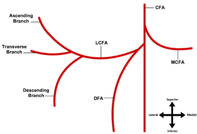
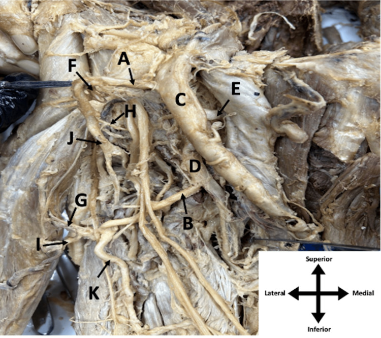
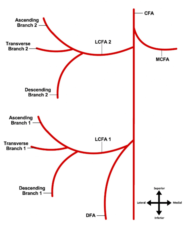
The lateral circumflex femoral artery (LCFA), a branch of the deep femoral artery (DFA), supplies the muscular and fascial anatomy of the anterior thigh. An undocumented variation of the LCFA was discovered during a dissection of the lower extremities. The LCFA is a vital vessel that can be used in coronary artery bypass grafts (CABGs) and reconstructive and bypass surgical procedures. On the other hand, the LCFA is susceptible to iatrogenic damage during surgeries involving the hip joint and procedures such as femoral nerve blocks. Knowledge of variations in the origin and course of the LFCA, like many other anatomical structures, is an important concept that physicians and health care providers must be aware of when performing anterior thigh procedures. This case report shows an interesting duplication of the LCFA, the first originating superiorly from the common femoral artery (CFA) and the second from the deep femoral artery (DFA) inferiorly. Both LCFAs exhibited typical trifurcation into ascending, descending, and transverse branches.
Keywords: anatomical variant; anatomy; cadaver; deep femoral artery; femoral artery; lateral circumflex femoral artery.
Copyright © 2024, Granger et al.
Conflict of interest statement
The authors have declared that no competing interests exist.
Figures
References
-
- Swift H, Bordoni B. Treasure Island (FL): StatPearls [Internet]; 2022. Anatomy, Bony Pelvis and Lower Limb: Femoral Artery. - PubMed
-
- Prough H, Launico MV, Alsayouri K. Anatomy, Bony Pelvis, and Lower Limb, Lateral Circumflex Femoral Artery. Treasure Island (FL): StatPearls [Internet]; 2022. - PubMed
-
- Arterial supply of sciatic nerve and its impact on clinical practice. Mohamed Metwally ESA, Aly El-Sekily NM, El Karim Ramadan NA. International Journal of Clinical and Developmental Anatomy. 2015;1:79–84.
-
- An analysis of the variations and clinical applications of the lateral circumflex femoral artery. Ma M, Sang H, Ye Y, et al. Folia Morphol (Warsz) 2021;80:557–566. - PubMed
Publication types
LinkOut - more resources
Full Text Sources