

Novel Surgical Approach for Large Intraosseous Subchondral Cysts of Talus: A Case Report and Technical Innovation
- PMID: 38344643
- PMCID: PMC10858398
- DOI: 10.7759/cureus.52078
Novel Surgical Approach for Large Intraosseous Subchondral Cysts of Talus: A Case Report and Technical Innovation
Abstract
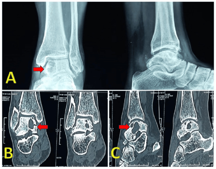
Large subchondral bone cysts in the medial talar body and dome are common and can cause persistent pain and swelling during axial loading. Open debridement and bone grafting are often necessary to treat these lesions but can require extensive soft-tissue dissection or malleolar osteotomies. A 40-year-old woman presented with ankle pain and swelling for 1 year, worsening with activity and no history of trauma. X-rays showed a cystic lesion in the medial talar dome with no joint line disruption. CT confirmed the cystic lesion without bone collapse or expansion. An anterior approach to the ankle joint was extended to access the talar neck. A window was created in the talar neck to debride and curette the medial talar dome, and the void was filled with allograft. The patient was non-weight-bearing for 6 weeks, followed by gradual weight-bearing and ankle range of motion exercises starting on postoperative day 1. The patient returned to her pre-injury status within 3 months and was asymptomatic at the 6-year follow-up, with good bone graft integration and no symptoms. This technical note presents a novel approach to lesions of the medial talar body and dome through the talar neck, avoiding the need for malleolar osteotomy or disruption to the tibiotalar joint, and resulting in good functional outcomes.
Keywords: bone grafting; intraosseous lesion; novel surgical approach; subchondral cyst; talar dome.
Copyright © 2024, Moonot et al.
Conflict of interest statement
The authors have declared that no competing interests exist.
Figures


References
-
- Primary aneurysmal bone cyst of talus. Sharma S, Gupta P, Sharma S, Singh M, Singh D. https://pubmed.ncbi.nlm.nih.gov/23853640/ J Res Med Sci. 2012;17:1192–1194. - PMC - PubMed
-
- Intra-osseous ganglion. Report of four cases and review of the literature. Pope TL Jr, Fechner RE, Keats TE. https://doi.org/10.1007/BF00360966. Skeletal Radiol. 1989;18:185–187. - PubMed
-
- Subchondral cysts of bone. Nigrisoli P, Beltrami P. Lo Scapello. 1971;1:65–75.
-
- Intraosseous ganglia: radioisotope bone imaging. Wise DI. https://pubmed.ncbi.nlm.nih.gov/1573612/ J R Coll Surg Edinb. 1992;37:57–58. - PubMed
-
- Massive intraosseous ganglion of the talus: reconstruction of the articular surface of the ankle joint. Koulalis D, Schultz W. https://doi.org/10.1053/jars.2000.8949 Arthroscopy. 2000;16:0. - PubMed
Publication types
LinkOut - more resources
Full Text Sources