

Structure Determination of Biogenic Crystals Directly from 3D Electron Diffraction Data
- PMID: 38344673
- PMCID: PMC10853906
- DOI: 10.1021/acs.cgd.3c01290
Structure Determination of Biogenic Crystals Directly from 3D Electron Diffraction Data
Abstract
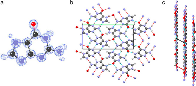
Highly reflective assemblies of purine, pteridine, and flavin crystals are used in the coloration and visual systems of many different animals. However, structure determination of biogenic crystals by single-crystal XRD is challenging due to the submicrometer size and beam sensitivity of the crystals, and powder XRD is inhibited due to the small volumes of powders, crystalline impurity phases, and significant preferred orientation. Consequently, the crystal structures of many biogenic materials remain unknown. Herein, we demonstrate that the 3D electron diffraction (3D ED) technique provides a powerful alternative approach, reporting the successful structure determination of biogenic guanine crystals (from spider integument, fish scales, and scallop eyes) from 3D ED data confirmed by analysis of powder XRD data. The results show that all biogenic guanine crystals studied are the previously known β-polymorph. This study highlights the considerable potential of 3D ED for elucidating the structures of biogenic molecular crystals in the nanometer-to-micrometer size range. This opens up an important opportunity in the development of organic biomineralization, for which structural knowledge is critical for understanding the optical functions of biogenic materials and their possible applications as sustainable, biocompatible optical materials.
© 2024 The Authors. Published by American Chemical Society.
Conflict of interest statement
The authors declare no competing financial interest.
Figures


Similar articles
-
Revealing the Crystal Structure of the Purine Base Xanthine with Three-Dimensional (3D) Electron Diffraction.Cryst Growth Des. 2025 Feb 11;25(5):1293-1298. doi: 10.1021/acs.cgd.4c01594. eCollection 2025 Mar 5. Cryst Growth Des. 2025. PMID: 40060986 Free PMC article.
-
Biogenic Guanine Crystals Are Solid Solutions of Guanine and Other Purine Metabolites.J Am Chem Soc. 2022 Mar 23;144(11):5180-5189. doi: 10.1021/jacs.2c00724. Epub 2022 Mar 7. J Am Chem Soc. 2022. PMID: 35255213 Free PMC article.
-
Application of X-ray Diffraction and Electron Crystallography for Solving Complex Structure Problems.Acc Chem Res. 2017 Nov 21;50(11):2737-2745. doi: 10.1021/acs.accounts.7b00366. Epub 2017 Nov 1. Acc Chem Res. 2017. PMID: 29091406
-
Single-crystal structure determination of nanosized metal-organic frameworks by three-dimensional electron diffraction.Nat Protoc. 2022 Oct;17(10):2389-2413. doi: 10.1038/s41596-022-00720-8. Epub 2022 Jul 27. Nat Protoc. 2022. PMID: 35896741 Review.
-
Three-dimensional electron diffraction as a complementary technique to powder X-ray diffraction for phase identification and structure solution of powders.IUCrJ. 2015 Feb 10;2(Pt 2):267-82. doi: 10.1107/S2052252514028188. eCollection 2015 Mar 1. IUCrJ. 2015. PMID: 25866663 Free PMC article. Review.
Cited by
-
Understanding the Solid-State Structure of Riboflavin through a Multitechnique Approach.Cryst Growth Des. 2024 Jul 18;24(15):6256-6266. doi: 10.1021/acs.cgd.4c00480. eCollection 2024 Aug 7. Cryst Growth Des. 2024. PMID: 39131447 Free PMC article.
-
Revealing the Crystal Structure of the Purine Base Xanthine with Three-Dimensional (3D) Electron Diffraction.Cryst Growth Des. 2025 Feb 11;25(5):1293-1298. doi: 10.1021/acs.cgd.4c01594. eCollection 2025 Mar 5. Cryst Growth Des. 2025. PMID: 40060986 Free PMC article.
-
Guanine Crystallization by Particle Attachment.J Am Chem Soc. 2025 Jun 4;147(22):19139-19147. doi: 10.1021/jacs.5c04543. Epub 2025 May 23. J Am Chem Soc. 2025. PMID: 40407389 Free PMC article.
-
MicroED: Unveiling the Structural Chemistry of Plant Biomineralisation.Molecules. 2024 Oct 17;29(20):4916. doi: 10.3390/molecules29204916. Molecules. 2024. PMID: 39459285 Free PMC article.
-
Rationalizing the Influence of Small-Molecule Dopants on Guanine Crystal Morphology.Chem Mater. 2024 Sep 1;36(18):8910-8919. doi: 10.1021/acs.chemmater.4c01771. eCollection 2024 Sep 24. Chem Mater. 2024. PMID: 39347467 Free PMC article.
References
-
- Gur D.; Palmer B. A.; Weiner S.; Addadi L. Light Manipulation by Guanine Crystals in Organisms: Biogenic Scatterers, Mirrors, Multilayer Reflectors and Photonic Crystals. Adv. Funct. Mater. 2017, 27 (6), 1603514.10.1002/adfm.201603514. - DOI
-
- Wagner A.; Wen Q.; Pinsk N.; Palmer B. A. Functional Molecular Crystals in Biology. Isr. J. Chem. 2021, 61 (9), 668–678. 10.1002/ijch.202100069. - DOI
-
- Levy-Lior A.; Pokroy B.; Levavi-Sivan B.; Leiserowitz L.; Weiner S.; Addadi L. Biogenic Guanine Crystals from the Skin of Fish May Be Designed to Enhance Light Reflectance. Cryst. Growth Des. 2008, 8 (2), 507–511. 10.1021/cg0704753. - DOI
LinkOut - more resources
Full Text Sources