Review
. 2024 Feb 27;24(5):1394-1418.
doi: 10.1039/d3lc00784g.

Microfluidic approaches in microbial ecology

Affiliations
Review

Microfluidic approaches in microbial ecology

Giovanni Stefano Ugolini et al. Lab Chip. .

Abstract

Microbial life is at the heart of many diverse environments and regulates most natural processes, from the functioning of animal organs to the cycling of global carbon. Yet, the study of microbial ecology is often limited by challenges in visualizing microbial processes and replicating the environmental conditions under which they unfold. Microfluidics operates at the characteristic scale at which microorganisms live and perform their functions, thus allowing for the observation and quantification of behaviors such as growth, motility, and responses to external cues, often with greater detail than classical techniques. By enabling a high degree of control in space and time of environmental conditions such as nutrient gradients, pH levels, and fluid flow patterns, microfluidics further provides the opportunity to study microbial processes in conditions that mimic the natural settings harboring microbial life. In this review, we describe how recent applications of microfluidic systems to microbial ecology have enriched our understanding of microbial life and microbial communities. We highlight discoveries enabled by microfluidic approaches ranging from single-cell behaviors to the functioning of multi-cellular communities, and we indicate potential future opportunities to use microfluidics to further advance our understanding of microbial processes and their implications.


Conflict of interest statement

The authors declare no conflicts of interest.

Figures

Fig. 1
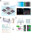
Fig. 1. Microfluidic systems used to quantify the response of individual bacteria to environmental fluctuations. (A) Diagram of the Mother Machine microfluidic chip, featuring a main channel for cell loading and media flow, and thousands of microchannels hosting single bacteria. Reproduced from ref. with permission from The American Association for the Advancement of Science, copyright 2018. (B) Sketches of the Mother Machine used to mimic a single environmental shift or periodical fluctuations. (C) Quantification of growth responses such as the lag time at single-cell level upon one nutrient switches applied in a Mother Machine. Reproduced from ref. with permission from National Academy of Sciences, copyright 2020. (D) Quantification of growth rates of cells exposed to periodic switches between high and low concentrations of nutrients. Reproduced from ref. with permission from Springer Nature, copyright 2021. (E) Transcriptional responses quantified using fluorescent reporters in microfluidics. Reproduced from ref. with permission from Wiley, copyright 2016. (F) Accumulation of auto-fluorescent compounds, such as nutrients and antibiotics, tracked in a Mother Machine. Reproduced from ref. and with permission from Springer Nature and RSC copyright 2020, 2022. (G) Sketches of the concept of transgeneration cellular memory. Microfluidics enables the dynamic tracking of fluorescence across cell lineages during environmental fluctuations, thus enabling the measurement of transgenerational cellular memory, in which cells in subsequent generations exposed to different environments retain similarities in the level of fluorescence-labeled functional proteins (left panel, blue circles). Similarly, transgenerational memory at the gene expression level (right panel, green circles) can be tracked, where cells maintain similarities in the expression levels of transcriptional reporters.
Fig. 2
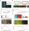
Fig. 2. Microfluidic approaches to study the effect of spatial heterogeneity on microorganisms. (A) Schematic of a source–sink device architecture. A central channel containing cells is flanked by two side channels where two different concentrations of a compound are flowed. Permeable side walls block the passage of cells but allow for transport and the formation of a defined (often linear) gradient within the central channel. (B) Effects of flow on bacteria at different locations in the water column: (i and ii) the torque from shear rotates cells or causes them to travel in spiral trajectories; (iii and iv) near a surface, cells can be oriented by shear in the direction opposite to the flow, causing upstream movement, either by flagellar propulsion or via the use of pili. (C) A microinjector geometry to study bacterial chemotaxis: a central channel is nested within a larger channel to form a nozzle. Nutrients are flowed through the nozzle to form a plume (red box, lower magnified view) within the larger channel hosting cells. Reproduced from ref. with permission from Wiley, copyright 2008. (D) Distribution of P. aeruginosa flowing around a micropillar. Non-motile cells preferentially attach to the windward side of the pillar, while motile cells are reoriented by shear and preferentially attach to the leeward side. Reproduced from ref. with permission from Springer Nature, copyright 2020. (E) Trapping of motile B. subtilis cells by shear. In these time series showing the spatial distribution of motile B. subtilis cells in a laminar flow, motile cells are rapidly depleted from the central low-shear regions when flow is applied, compared to experiments with no flow applied or with flow applied to dead cells. Reproduced from ref. with permission from Springer Nature, copyright 2014.
Fig. 3
Fig. 3. Schematic overview of typical microfluidic systems for studying microbial interactions in clonal populations. (A) Left: A Family Machine, containing one main channel and multiple side microchambers to culture bacteria monolayers. Right: Representative applications of the Family Machine setup including to establish nutrient gradients through the chamber to quantify the resulting metabolic heterogeneity of a population, or to evaluate aggregation behavior of cell populations. (B) The Mother Machine device connected to an external flask hosting a pair of interacting bacteria enables flow of the medium from such interactions to the same strains cultured in the Mother Machine, thus monitoring single-cell growth rates in response to the evolution of the interaction in the flask. Reproduced from ref. with permission from Springer Nature, copyright 2023. (C) A Family Machine containing two main channels has been used to investigate contact-dependent interactions. When this device was used to investigate T6SS-mediated killing, “target” cells (purple) were inoculated into the center of the chamber, followed by inoculation of “attacker” cells (green) at the edges of the chamber. This spatial arrangement enables time-lapse tracking of the boundary between cells of the two species, where the interaction occurs. Reproduced from ref. with permission from PLOS, copyright 2020.
Fig. 4
Fig. 4. Microfluidic approaches to quantify the interaction distance among microbial cells. (A) Using microfluidics, populations can be spatially isolated within separate chambers using a barrier that prevents the passage of cells while permitting the exchange of compounds. In this way, the interaction distance can be quantified by determining the farthest point from the barrier at which cross-fed cells are able to grow. Reproduced from ref. with permission from RSC, copyright 2019. (B) The MISTiC microfluidics design allows direct measurement of how the spatial distance between sender and receiver cell populations affects their interactions mediated by diffusible metabolites, by introducing a diffusion chamber of varying length between the two populations of cells. Reproduced from ref. with permission from Springer Nature, copyright 2020. (C and D) Genetically distinct populations often form spatially segregated cell clusters arising from different founder cells within a microfluidic chamber. Cell clusters can be differentiated using fluorescent labeling. (C) In the case of metabolic exchange of amino acids, the interaction range has been quantified by correlating the growth rates of individual focal cells with the fraction of the community within a given neighborhood distance that is constituted by the interacting partner. The interaction range is then defined as the neighborhood distance with maximal correlation coefficient. Reproduced from ref. with permission from Springer Nature, copyright 2020. (D) In the case of signaling between different cell populations, the interaction range can be quantified as the maximal distance from the boundary at which receiver cells show fluorescent response. Reproduced from ref. with permission from Springer Nature, copyright 2021.
Fig. 5
Fig. 5. Applications of microfluidics in biofilm research. (A) Surface-attached biofilms in a microfluidic channel can be exposed to a controlled flow, impacting nutrient and oxygen concentration, quorum sensing, and exerting hydrodynamic forces on the superficial layer. (B) Flow modulates cell structuring in surface-attached biofilms. Flow rate determines the cell patterning of two fluorescently labeled C. crescentus populations within a straight microchannel, resulting in limited clonal segregation at a low flow rate (left) and scattered monoclonal colonies at a high flow rate (right). Reproduced from ref. with permission from Springer Nature, copyright 2019. (C) Streamers are biofilm filaments surrounded by fluid flow. Secondary flow around the curved surfaces of a pillar causes biomass to accumulate in the midplane, while the downstream flow extrudes it downstream of the pillar. (D) Streamers formed by Sagittula stellata on a single oil droplet in a microfluidic channel. Reproduced from ref. with permission from Springer Nature, copyright 2020. (E) Activation of quorum sensing in a thick Staphylococcus aureus biofilm exposed to flow in a microfluidic channel (upper panel). Lower panels: Comparison of the base of biofilms grown at low (left) and high flow rates (right). Quorum sensing is active in yellow cells, inactive in red cells. Reproduced from ref. with permission from Springer Nature, copyright 2016. (F) The diffuse mixer microfluidic channel dispenses quorum sensing signaling molecules and two dispersal proteins during biofilm growth. Reproduced from ref. with permission from Springer Nature, copyright 2012.

References

    1. Lozupone C. A. Stombaugh J. I. Gordon J. I. Jansson J. K. Knight R. Diversity, stability and resilience of the human gut microbiota. Nature. 2012;489(7415):220–230. doi: 10.1038/nature11550. - DOI - PMC - PubMed
    1. Martin-Platero A. M. Cleary B. Kauffman K. Preheim S. P. McGillicuddy D. J. Alm E. J. Polz M. F. High resolution time series reveals cohesive but short-lived communities in coastal plankton. Nat. Commun. 2018;9(1):266. doi: 10.1038/s41467-017-02571-4. - DOI - PMC - PubMed
    1. Tinta T. Vojvoda J. Mozeticč P. Talaber I. Vodopivec M. Malfatti F. Turk V. Bacterial community shift is induced by dynamic environmental parameters in a changing coastal ecosystem (northern Adriatic, northeastern Mediterranean Sea) – a 2-year time-series study. Environ. Microbiol. 2014;17(10):3581–3596. doi: 10.1111/1462-2920.12519. - DOI - PubMed
    1. Mancuso C. P. Lee H. Abreu C. I. Gore J. Khalil A. S. Environmental fluctuations reshape an unexpected diversity-disturbance relationship in a microbial community. eLife. 2021;10:e67175. doi: 10.7554/eLife.67175. - DOI - PMC - PubMed
    1. Nguyen J. Lara-Gutiérrez J. Stocker R. Environmental fluctuations and their effects on microbial communities, populations and individuals. FEMS Microbiol. Rev. 2020;45(4):fuaa068. doi: 10.1093/femsre/fuaa068. - DOI - PMC - PubMed

Publication types