. 2024 Feb 20;96(7):3077-3086.
doi: 10.1021/acs.analchem.3c05194. Epub 2024 Feb 12.

Deuterium Labeling of Isoaspartic and Isoglutamic Acids for Mass Spectrometry Analysis

Affiliations

Deuterium Labeling of Isoaspartic and Isoglutamic Acids for Mass Spectrometry Analysis

Masaru Miyagi et al. Anal Chem. .

Abstract

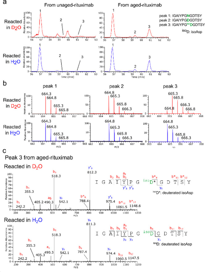
Isoaspartic acid (isoAsp) is a common protein modification that spontaneously arises from asparagine or aspartic acid and has been linked to various diseases and health conditions. However, current methods for identifying isoAsp sites in proteins often suffer from ambiguity and have not gained widespread adoption. We developed a novel method that exclusively labels isoAsp with deuterium. This method capitalizes on the unique structural characteristics of isoAsp residues, which possess a free α-carboxyl group and can form an oxazolone ring. Once the oxazolone ring forms, it facilitates racemization at the Cα-position, incorporating a deuteron from a D2O solvent. The sites of deuterium-incorporated isoAsp in proteins can be unequivocally determined by comparing the precursor and product ion masses of the peptides from proteins reacted in H2O and D2O. The effectiveness of this method has been demonstrated through its application to model proteins lysozyme and rituximab. Furthermore, we have confirmed that the isoAsp deuterium-labeling reaction efficiently labels both l- and d-isoAsp without distinction, as well as isoglutamic acid (isoGlu), for which no effective detection methods currently exist.


Conflict of interest statement

The authors declare no competing financial interest.

Figures

Scheme 1
Scheme 1. Pathways for Spontaneous Deamidation, Isomerization, and Racemization of l-Asn and l-Asp Residues in Proteins
Deamidation of l-Asn begins with the backbone nitrogen of the C-terminal neighboring residue attacking the side-chain carbonyl carbon of Asn, forming succinimide intermediate I. Hydrolysis of intermediate I leads to l-Asp or l-isoAsp. The intermediate I can also undergo racemization via its enol form II, which produces the D configuration of succinimide intermediate III. Hydrolysis of intermediate III yields d-Asp or d-isoAsp. Isomerization and racemization of l-Asp begin similarly with the backbone nitrogen of the C-terminal neighboring residue attacking the side-chain carbonyl carbon of Asp, forming succinimide intermediate I. Hydrolysis of intermediate I leads to either the original l-Asp or l-isoAsp. l-Asp can also be converted to d-isoAsp or d-Asp via intermediates II and III as l-Asn.
Scheme 2
Scheme 2. Deuterium Labeling of isoAsp and isoGlu
In the presence of pyridine and acetic anhydride, acetylpyridinium is formed, which then reacts with the α-carboxyl group of isoX (X = Asp when n = 1; X = Glu when n = 2) to form mixed anhydride IV. The α-hydrogen (α-H) of VI in the keto form is abstracted by pyridine in step a1 to form an enolate anion represented by two resonance hybrids VII and VIII. The subsequent reaction of this enolate with deuterium (red) in D2O in step a2 gives a racemic oxazolone VIV incorporating a deuteron at the α-position. The yield of deuterium labeling is significantly enhanced by the repetitive process of generating an oxazolone VI from IsoX through step b, as long as a large excess of acetic anhydride remains in the reaction mixture. The labile amide, hydroxy, and carboxy protons (blue) being exchanged with deuterons in D2O are exchanged back to protons in H2O, but the deuterons incorporated into the α-position of isoX remain unexchanged.
Figure 1
Figure 1
(a) LC–MS analysis of isoDG, DG, and NG subjected to the deuterium-labeling reaction. Each peptide was dissolved in 40 μL of D2O/pyridine (1:3, v/v) or H2O/pyridine (1:3, v/v) and reacted with 30 μL of acetic anhydride (Ac2O) at room temperature for 30 min. After the reaction, the peptide was dried and further incubated in 50 μL of a hydrazine solution (0.35 wt % in H2O) at room temperature for 30 min. The resulting peptide was then analyzed by LC–MS. The total ion chromatograms (m/z 300–1800) and mass spectra of the three peptides reacted in D2O (red) or H2O (blue) are shown. (b) MS/MS spectra of isoDG reacted in D2O (upper MS/MS spectrum, precursor ions: m/z 663.3, z = 1) or H2O (lower MS/MS spectrum, precursor ions: m/z 662.3, z = 1). The fragment ions that contain a deuterated isoAsp residue are indicated with an asterisk.
Figure 2
Figure 2
LC–MS/MS analyses of aged and unaged lysozyme subjected to the predigestion isoAsp-labeling reaction. Aged and unaged lysozymes were dissolved in 120 μL of D2O/pyridine (1:3, v/v) or H2O/pyridine (1:3, v/v) and reacted with 90 μL of acetic anhydride (Ac2O) at room temperature for 30 min. After the reaction, the mixture was dried and further incubated in 50 μL of a hydrazine solution (0.35 wt % in H2O) at room temperature for 30 min. Subsequently, the protein was digested by chymotrypsin and analyzed by LC–MS/MS. (a) Extracted ion chromatograms (m/z 875.89–877.91) encompassing the doubly charged ions of the following three peptides: peak 1: CAKKIVSDGNGMNAW, peak 2: CAKKIVSDGDGMNAW, and peak 3: CAKKIVSDGisoDGMNAW. (b) Mass spectra of peaks 1–3 from the lysozyme reacted in D2O or H2O. (c) MS/MS spectra of peak 3 from the aged lysozyme reacted in D2O or H2O. The fragment ions incorporating a deuterium atom are indicated with an asterisk.
Figure 3
Figure 3
Representative LC–MS/MS data for one of the isoAsp sites identified in rituximab. Aged and unaged rituximab were dissolved in 120 μL of D2O/pyridine (1:3, v/v) or H2O/pyridine (1:3, v/v) and reacted with 90 μL of acetic anhydride (Ac2O) at room temperature for 30 min. After the reaction, the mixture was dried and further incubated in 50 μL of a hydrazine solution (0.35 wt % in H2O) at room temperature for 30 min. Subsequently, the protein was digested by chymotrypsin and analyzed by LC–MS/MS. (a) Extracted ion chromatograms (m/z 664.29–666.31) encompassing the doubly charged ions of the following three peptides containing amino acid residue 55 of the heavy chain of rituximab: peak 1: IGAIYPGNGDTSY, peak 2: IGAIYPGDGDTSY, and peak 3: IGAIYPGisoDGDTSY. Residue 55 is underlined. (b) Mass spectra of peaks 1–3 from rituximab reacted in D2O or H2O. (c) MS/MS spectra of peak 3 from aged rituximab reacted in D2O or H2O. The fragment ions incorporating a deuterium atom are indicated with an asterisk.
Figure 4
Figure 4
(a) LC–MS analysis of isoEG, EG, and QG peptides subjected to the deuterium-labeling reaction. Each peptide was dissolved in 40 μL of D2O/pyridine (1:3, v/v) or H2O/pyridine (1:3, v/v) and reacted with 30 μL of acetic anhydride (Ac2O) at room temperature for 30 min. After the reaction, the peptide was dried and further incubated in 50 μL of a hydrazine solution (0.35 wt % in H2O) at room temperature for 30 min. The resulting peptide was then analyzed by LC–MS. The total ion chromatograms (m/z 300–1800) and mass spectra of the three peptides reacted in D2O (red) or H2O (blue) are shown. (b) MS/MS spectra of isoEG reacted in D2O (upper MS/MS spectrum, precursor ions: m/z 773.3, z = 1) or H2O (lower MS/MS spectrum, precursor ions: m/z 772.3, z = 1). The m/z values of the fragment ions containing deuterated isoGlu residues in the upper spectrum are indicated with an asterisk.

References

    1. Geiger T.; Clarke S. Deamidation, isomerization, and racemization at asparaginyl and aspartyl residues in peptides. Succinimide-linked reactions that contribute to protein degradation. J. Biol. Chem. 1987, 262, 785–794. 10.1016/S0021-9258(19)75855-4. - DOI - PubMed
    1. Stephenson R. C.; Clarke S. Succinimide Formation from Aspartyl and Asparaginyl Peptides as a Model for the Spontaneous Degradation of Proteins. J. Biol. Chem. 1989, 264, 6164–6170. 10.1016/S0021-9258(18)83327-0. - DOI - PubMed
    1. Roher A. E.; Lowenson J. D.; Clarke S.; Wolkow C.; Wang R.; Cotter R. J.; Reardon I. M.; Zurcher-Neely H. A.; Heinrikson R. L.; Ball M. J.; Greenberg B. D. Structural alterations in the peptide backbone of beta-amyloid core protein may account for its deposition and stability in Alzheimer’s disease. J. Biol. Chem. 1993, 268, 3072–3083. 10.1016/s0021-9258(18)53661-9. - DOI - PubMed
    1. Watanabe A.; Takio K.; Ihara Y. Deamidation and Isoaspartate Formation in Smeared Tau in Paired Helical Filaments. J. Biol. Chem. 1999, 274, 7368–7378. 10.1074/jbc.274.11.7368. - DOI - PubMed
    1. Nowak C.; Cheung J. M.; M Dellatore S.; Katiyar A.; Bhat R.; Sun J.; Ponniah G.; Neill A.; Mason B.; Beck A.; Liu H. Forced degradation of recombinant monoclonal antibodies: A practical guide. mAbs 2017, 9, 1217–1230. 10.1080/19420862.2017.1368602. - DOI - PMC - PubMed

Publication types