

Transcript Profiles of Microglia/Macrophage Cells at the Borders of Chronic Active and Subpial Gray Matter Lesions in Multiple Sclerosis
- PMID: 38345145
- PMCID: PMC11060930
- DOI: 10.1002/ana.26877
Transcript Profiles of Microglia/Macrophage Cells at the Borders of Chronic Active and Subpial Gray Matter Lesions in Multiple Sclerosis
Abstract
Objective: Microglia/macrophages line the border of demyelinated lesions in both cerebral white matter and the cortex in the brains of multiple sclerosis patients. Microglia/macrophages associated with chronic white matter lesions are thought to be responsible for slow lesion expansion and disability progression in progressive multiple sclerosis, whereas those lining gray matter lesions are less studied. Profiling these microglia/macrophages could help to focus therapies on genes or pathways specific to lesion expansion and disease progression.
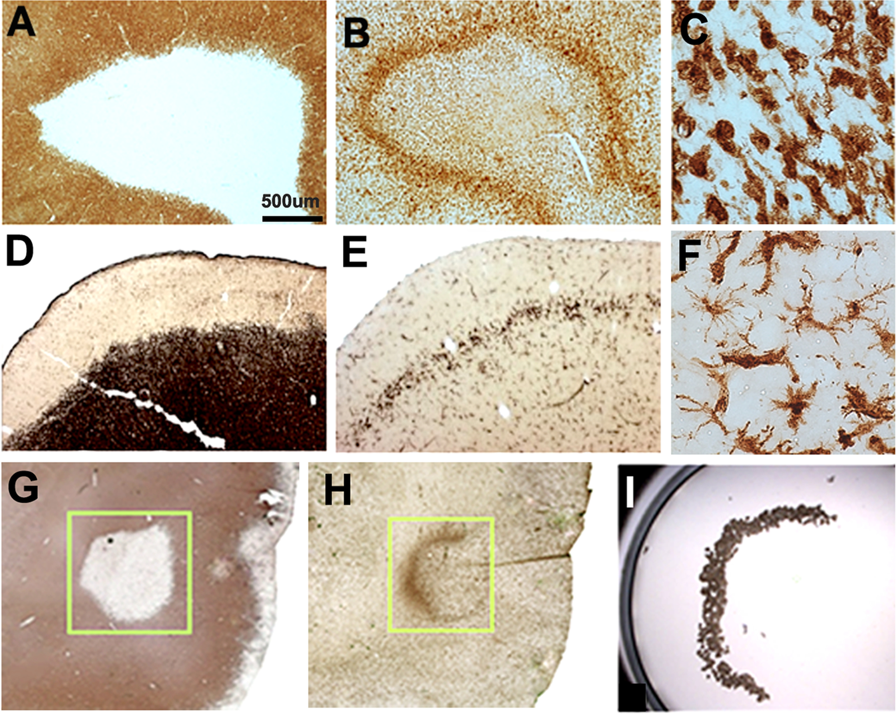
Methods: We compared the morphology and transcript profiles of microglia/macrophages associated with borders of white matter (WM line) and subpial gray matter lesions (GM line) using laser capture microscopy. We performed RNA sequencing on isolated cells followed by immunocytochemistry to determine the distribution of translational products of transcripts increased in WM line microglia.
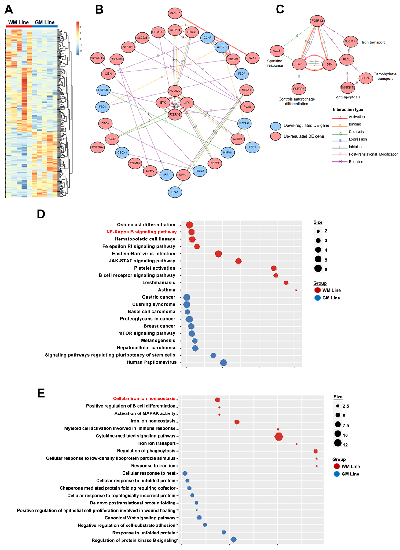
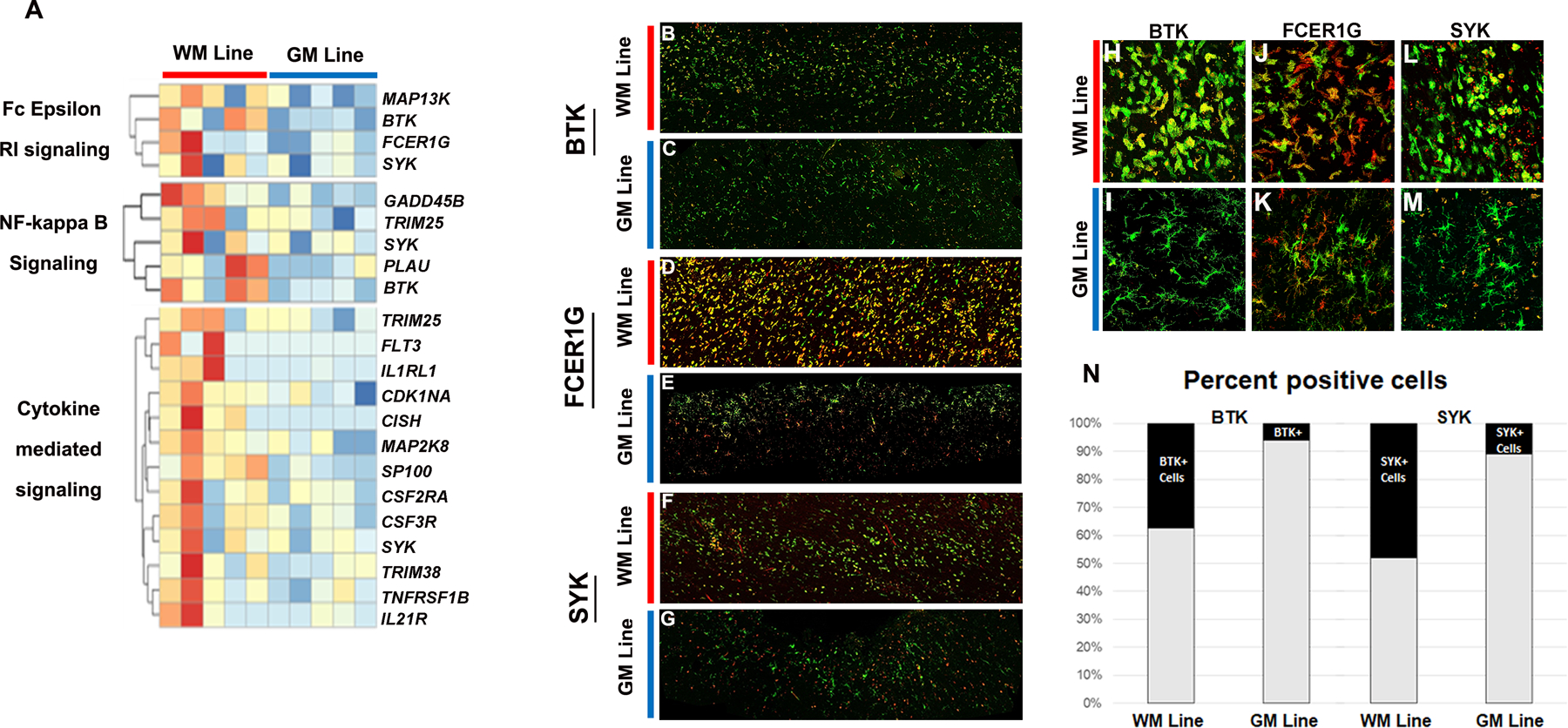
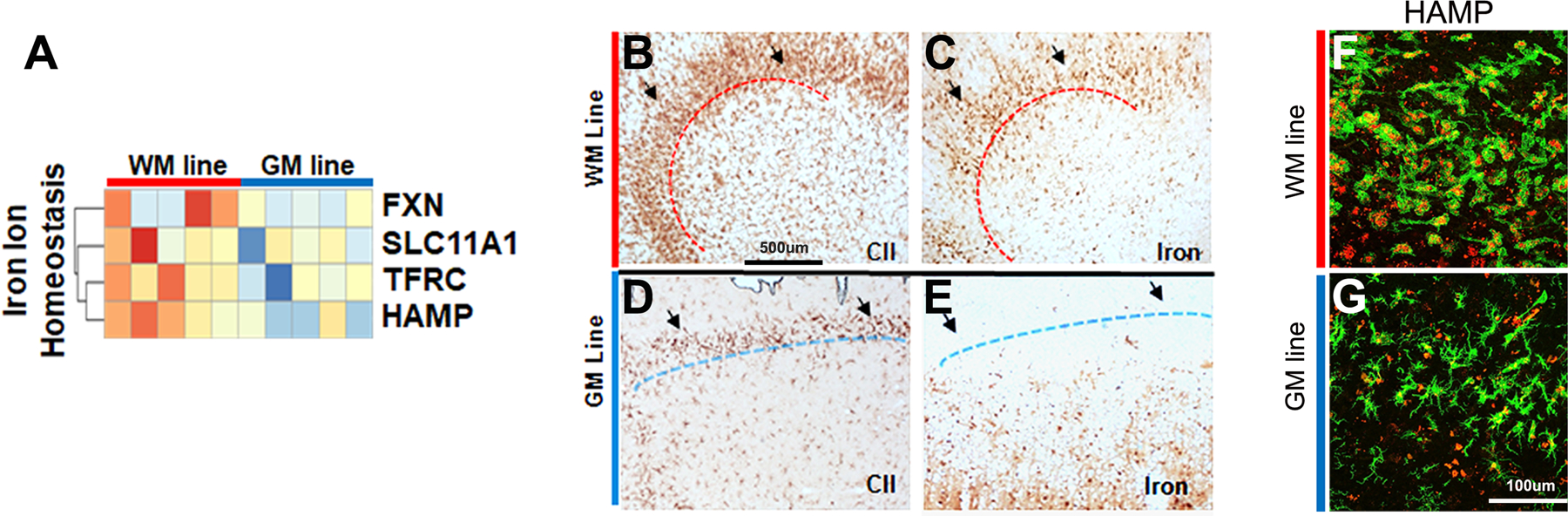
Results: Cells in the WM line appear activated, with shorter processes and larger cell bodies, whereas those in the GM line appear more homeostatic, with smaller cell bodies and multiple thin processes. Transcript profiling revealed 176 genes in WM lines and 111 genes in GM lines as differentially expressed. Transcripts associated with immune activation and iron homeostasis were increased in WM line microglia, whereas genes belonging to the canonical Wnt signaling pathway were increased in GM line microglia.
Interpretation: We propose that the mechanisms of demyelination and dynamics of lesion expansion are responsible for differential transcript expression in WM lines and GM lines, and posit that increased expression of the Fc epsilon receptor, spleen tyrosine kinase, and Bruton's tyrosine kinase, play a key role in regulating microglia/macrophage function at the border of chronic active white matter lesions. ANN NEUROL 2024;95:907-916.
© 2024 The Authors. Annals of Neurology published by Wiley Periodicals LLC on behalf of American Neurological Association.
Conflict of interest statement
Potential Conflicts of Interest
The authors report no conflicts of interest.
Figures
Similar articles
-
Regulation of microglial TMEM119 and P2RY12 immunoreactivity in multiple sclerosis white and grey matter lesions is dependent on their inflammatory environment.Acta Neuropathol Commun. 2019 Dec 11;7(1):206. doi: 10.1186/s40478-019-0850-z. Acta Neuropathol Commun. 2019. PMID: 31829283 Free PMC article.
-
Transcriptional profiling of human microglia reveals grey-white matter heterogeneity and multiple sclerosis-associated changes.Nat Commun. 2019 Mar 13;10(1):1139. doi: 10.1038/s41467-019-08976-7. Nat Commun. 2019. PMID: 30867424 Free PMC article.
-
Small heat shock proteins are induced during multiple sclerosis lesion development in white but not grey matter.Acta Neuropathol Commun. 2015 Dec 22;3:87. doi: 10.1186/s40478-015-0267-2. Acta Neuropathol Commun. 2015. PMID: 26694816 Free PMC article.
-
Relationship Between White Matter Lesions and Gray Matter Atrophy in Multiple Sclerosis: A Systematic Review.Neurology. 2022 Apr 12;98(15):e1562-e1573. doi: 10.1212/WNL.0000000000200006. Epub 2022 Feb 16. Neurology. 2022. PMID: 35173016 Free PMC article.
-
MicroRNAs in gray and white matter multiple sclerosis lesions: impact on pathophysiology.J Pathol. 2020 Apr;250(5):496-509. doi: 10.1002/path.5399. Epub 2020 Mar 5. J Pathol. 2020. PMID: 32073139 Review.
Cited by
-
Recent developments in multiple sclerosis neuropathology.Curr Opin Neurol. 2025 Jun 1;38(3):173-179. doi: 10.1097/WCO.0000000000001370. Epub 2025 Apr 3. Curr Opin Neurol. 2025. PMID: 40178490 Free PMC article. Review.
-
The role of microglia in multiple sclerosis: implications for treatment with Bruton's tyrosine kinase inhibitors.Front Immunol. 2025 May 15;16:1495529. doi: 10.3389/fimmu.2025.1495529. eCollection 2025. Front Immunol. 2025. PMID: 40443664 Free PMC article. Review.
-
Pathways to Progressive Disability in Multiple Sclerosis: The Role of Glial Cells in Chronic CNS Inflammation.Glia. 2025 Oct;73(10):1928-1950. doi: 10.1002/glia.70044. Epub 2025 May 23. Glia. 2025. PMID: 40406903 Free PMC article. Review.
-
Inflammation alters myeloid cell and oligodendroglial iron-handling in multiple sclerosis.Acta Neuropathol Commun. 2025 Jun 4;13(1):124. doi: 10.1186/s40478-025-02020-0. Acta Neuropathol Commun. 2025. PMID: 40468400 Free PMC article.
-
Imaging Outcomes for Phase 2 Trials Targeting Compartmentalized Inflammation.Mult Scler. 2024 Dec;30(5_suppl):48-60. doi: 10.1177/13524585241301303. Mult Scler. 2024. PMID: 39658905 Free PMC article. Review.
References
-
- Healy LM, Stratton JA, Kuhlmann T, Antel J. The role of glial cells in multiple sclerosis disease progression. Nat. Rev. Neurol 2022;18:237–248. - PubMed
-
- Calvi A, Carrasco FP, Tur C et al. Association of Slowly Expanding Lesions on MRI With Disability in People With Secondary Progressive Multiple Sclerosis. Neurology 2022; - PubMed
Publication types
MeSH terms
Grants and funding
LinkOut - more resources
Full Text Sources
Medical
