. 2024 Jun;13(14):e2301966.
doi: 10.1002/adhm.202301966. Epub 2024 Feb 25.

Neutrophil Granulopoiesis Optimized Through Ex Vivo Expansion of Hematopoietic Progenitors in Engineered 3D Gelatin Methacrylate Hydrogels

Affiliations

Neutrophil Granulopoiesis Optimized Through Ex Vivo Expansion of Hematopoietic Progenitors in Engineered 3D Gelatin Methacrylate Hydrogels

Evan Cirves et al. Adv Healthc Mater. 2024 Jun.

Abstract

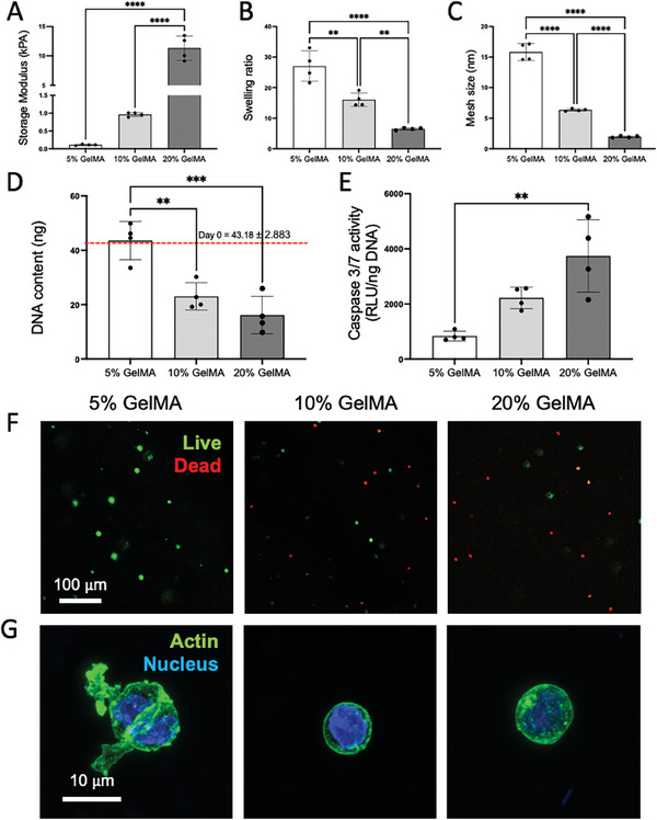
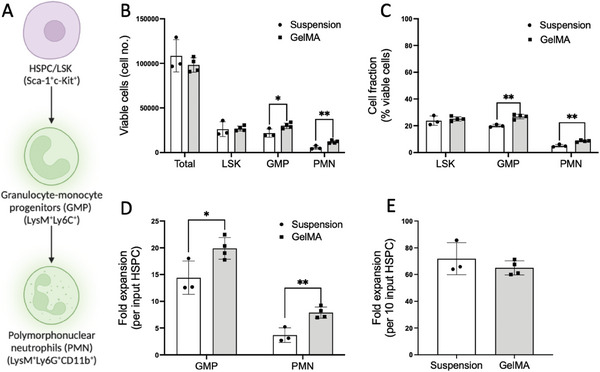
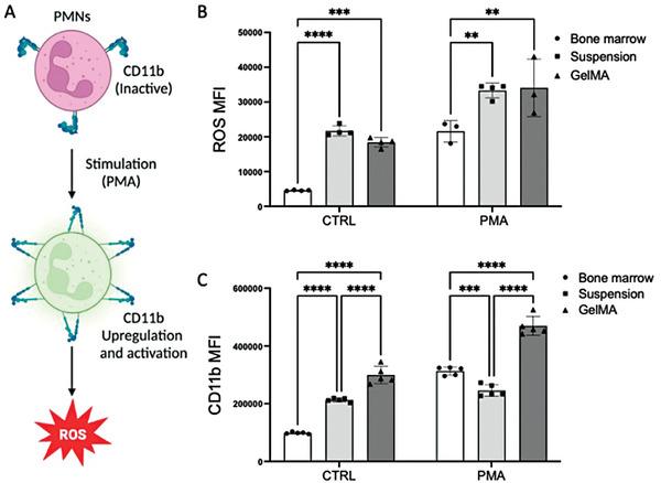
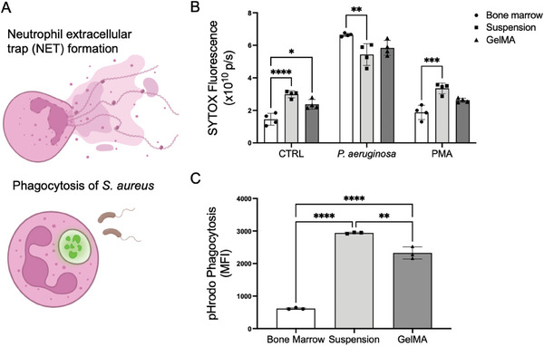
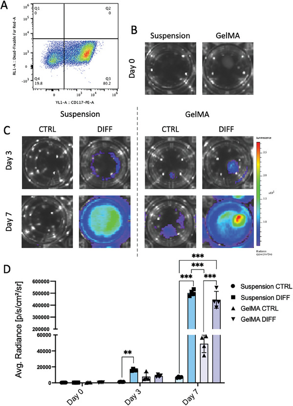
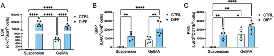
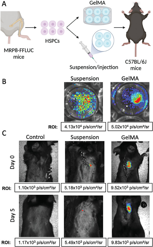
Neutrophils are the first line of defense of the innate immune system. In response to methicillin-resistant Staphylococcus aureus infection in the skin, hematopoietic stem, and progenitor cells (HSPCs) traffic to wounds and undergo extramedullary granulopoiesis, producing neutrophils necessary to resolve the infection. This prompted the engineering of a gelatin methacrylate (GelMA) hydrogel that encapsulates HSPCs within a matrix amenable to subcutaneous delivery. The authors study the influence of hydrogel mechanical properties to produce an artificial niche for granulocyte-monocyte progenitors (GMPs) to efficiently expand into functional neutrophils that can populate infected tissue. Lin-cKIT+ HSPCs, harvested from fluorescent neutrophil reporter mice, are encapsulated in GelMA hydrogels of varying polymer concentration and UV-crosslinked to produce HSPC-laden gels of specific stiffness and mesh sizes. Softer 5% GelMA gels yield the most viable progenitors and effective cell-matrix interactions. Compared to suspension culture, 5% GelMA results in a twofold expansion of mature neutrophils that retain antimicrobial functions including degranulation, phagocytosis, and ROS production. When implanted dermally in C57BL/6J mice, luciferase-expressing neutrophils expanded in GelMA hydrogels are visualized at the site of implantation for over 5 days. They demonstrate the potential of GelMA hydrogels for delivering HSPCs directly to the site of skin infection to promote local granulopoiesis.

Keywords: extramedullary granulopoiesis; gelatin methacrylate (GelMA); hematopoietic stem and progenitor cells (HSPCs); neutrophils.


Conflict of interest statement

The authors declare no conflict of interest.

Figures

Figure 1
Figure 1
GelMA hydrogel mechanical properties correlate with HSPC survival and a motile morphology. A) Storage modulus, B) swelling ratio, and C) mesh size of either 5%, 10%, or 20% GelMA hydrogels after production (N = 4). D) DNA content and E) caspase 3/7 activity in HSPC‐laden GelMA hydrogels 24 h after cell encapsulation (N = 4). F) Live/dead (living cells in green, dead cells in red) characterization of encapsulated HSPCs 24 h after hydrogel production. G) Fluorescent imaging of HSPC actin cytoskeleton in the different GelMA gels 24 h after production. For F and G, representative images out of six, two independent experiments. ** Denotes significance (p < 0.01); **** (p < 0.0001).
Figure 2
Figure 2
HSPC encapsulation into 5% GelMA hydrogels increases their neutrophil differentiation potential in comparison to suspension culture. A) To assess the expansion potential of HSPCs toward neutrophils, LysM‐EGFP cells were stained with Sca‐1+c‐KIT+ to determine undifferentiated HSPCs (LSK cells), with Ly6C+ to determine granulocyte‐monocyte progenitors (GMPs), and with Ly6G+CD11b+ to assess the polymorphonuclear neutrophil populations (PMNs). After 72 h of in vitro culture in either suspension or 5% GelMA gels, cells were isolated, and the B) total number of viable cells was determined through FACS. C) Cell fractions (% of viable cells) for LSK, GMP, and PMN cells were also determined. D) Myeloid expansion per input HSPC toward GMP or PMN phenotypes and E) the total fold expansion of all cells detected per input HSPCs were also determined after the culture time. N = 3 in suspension and 4 in GelMA. Suspension data points are represented with a dot symbol and white bars, and GelMA with a square symbol and light grey bars. * Denotes significance (p < 0.05) in comparison to the rest of the groups; ** (p < 0.01) in comparison to the rest of the groups.
Figure 3
Figure 3
Phenotypic differences between HSPCs that escaped the GelMA hydrogels and cells that remained inside the biomaterial after 7 days of in vitro culture in differentiation conditions. A) Graphical representation of analyzed cell populations defining the cells that remained encapsulated in the GelMA gels (In‐GelMA) versus cells that escaped the hydrogel (Escaped). B) Percentage of LSK, GMP, and PMN cells in suspension or GelMA (Escaped + In‐GelMA cells) after 7 days. D) Percentage of LSK, GMP, and PMN cells in suspension, in‐GelMA, and escaped after 7 days of differentiation culture. E) Median Fluorescence Intensity (MFI) of LysM‐EGP+ cells after 7 days of differentiation culture. N = 3 for all experiments. Suspension data points are represented with a dot symbol and white bars, GelMA with a square symbol and light grey bars, and Escaped with a triangle symbol and dark grey bars. * Denotes significance (p < 0.05) in comparison to the rest of the groups; ** (p < 0.01).
Figure 4
Figure 4
Analysis of ROS production and CD11b expression in neutrophils expanded from HSPCs encapsulated in GelMA compared to suspension‐grown and bone marrow isolated. A) Schematic showing inactivated neutrophils responding to PMA and subsequently upregulating and activating CD11b and increasing ROS production. B) Reactive Oxygen Species (ROS) Median Fluorescence Intensity (MFI) of CD11b+ neutrophils extracted from bone marrow (C57BL/6J mice) or cultured in vitro for 7 days in either suspension or GelMA, when exposed to no stimulus (CTRL) or to 10 ng mL−1 of PMA. C) CD11b MFI in CD11b+/LysM+ neutrophils extracted from the bone marrow (LysM‐EGFP mice) or cultured in vitro for 7 days in either suspension or GelMA, N = 3–5 for both experiments. Bone marrow data points are represented with a dot symbol and white bars, Suspension with a square symbol and light grey bars, and GelMA with a triangle symbol and dark grey bars. ** Denotes significance (p < 0.01), *** (p < 0.001), and **** (p < 0.0001).
Figure 5
Figure 5
Analysis of neutrophil function in response to bacterial infection. A) Schematic defining the biological responses measured in this figure. B) Average fluorescence of SYTOX Orange as an analog for NETosis. C) Average fluorescence pH‐reactive S. aureus bioparticles phagocytosed by EGFP+ cells and measured via AttuneNxT flow cytometer. Cells in both experiments were seeded and expanded for 7 days. N = 3–4. * Denotes significance (p < 0.05), ** (p < 0.01), *** (p < 0.001), and **** (p < 0.0001).
Figure 6
Figure 6
Luciferase activity of MRP8‐FFLUC HSPCs cultured in vitro in either suspension or encapsulated in 5% GelMA hydrogels. A) Confirmation of >80% of CD117+ HSPCs after cell isolation from the bone marrow of MRP8‐FFLUC mice. B) Luciferase activity at day 0 after isolation in cells in suspension or encapsulated in 5% GelMA gels. C) Luciferase activity after 3 and 7 days of in vitro culture in cells cultured in suspension or encapsulated in GelMA gels and maintained in basal (CTRL) or differentiation (DIFF) media. D) Quantification of luciferase activity (N = 4). Suspension CTRL data points are represented with a dot symbol and light blue bars, Suspension Diff with a square symbol and darker blue, GelMA CTRL with a triangle and light purple bars, and GelMA Diff with an inverted triangle and darker purple bars. ** Denotes significance (p < 0.01) and *** (p < 0.001).
Figure 7
Figure 7
Phenotypic characterization of MRP8‐FFLUC cell populations cultured in suspension or GelMA in maintenance (CTRL) or differentiation (DIFF) media. (A‐C) Number of LSK (Sca‐1+c‐kit+), GMP (Ly6C+c‐kit+), and PMN (Ly6G+CD11b+) cells after 7 days of in vitro culture. N = 5 for all experiments. CTRL and DIFF data points are represented with dot symbols and clear blue bars and with square symbols and dark blue bars respectively. * Denotes significance (p < 0.05); ** (p < 0.01) and **** (p < 0.0001).
Figure 8
Figure 8
Transplantation of neutrophils expanded from MRP8‐FFLUC HSPCs cultured in either suspension or GelMA gels into C57BL/6J mice. A) Schematic of the experimental flow. B) Luciferase activity in the suspension and GelMA groups after 7 days of in vitro culture. C) Luciferase activity of cells in suspension or GelMA at days 0 and 5 after transplantation into C57BL/6J mice.

Similar articles

References

    1. Mölne L., Verdrengh M., Tarkowski A., Infect. Immun. 2000, 68, 6162. - PMC - PubMed
    1. Verdrengh M., Tarkowski A., Infect. Immun. 1997, 65, 2517. - PMC - PubMed
    1. Hirai H., Zhang P., Dayaram T., Hetherington C. J., Mizuno S., Imanishi J., Akashi K., Tenen D. G., Nat. Immunol. 2006, 7, 732. - PubMed
    1. Nauseef W. M., Borregaard N., Nat. Immunol. 2014, 15, 602. - PubMed
    1. Hämäläinen S., Kuittinen T., Matinlauri I., Nousiainen T., Koivula I., Jantunen E., Leuk Lymphoma 2008, 49, 495. - PubMed

Publication types