

Quantitative and qualitative 3D analysis of mandibular lingual concavities: Implications for dental implant planning in the posterior mandible
- PMID: 38345362
- PMCID: PMC10860544
- DOI: 10.1002/cre2.858
Quantitative and qualitative 3D analysis of mandibular lingual concavities: Implications for dental implant planning in the posterior mandible
Abstract
Objective: The purpose of this study is to investigate the type of ridge (degree of angulation of the lingual concavity) and the buccolingual dimensions in the area of the first and second molars in both genders of different ages and how this will affect implant placement in the posterior mandible.
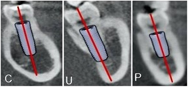
Materials and methods: This retrospective cross-sectional study comprised cone beam computed tomography images of 150 dental patients (75 males and 71 aged ≥30). The following were measured/reported: type (morphology) of the ridge (convex [C], parallel [P], or undercut [U]), buccolingual width at the base and the crest of the ridge, and ridge height. The concavity angle, depth, and length of the U-shaped ridge were measured too.
Results: The prevalence of type U ridge ranged from 32.7% in the first molar region to 62.7% in the second molar region. Almost all measurements and ridge type distributions were comparable amongst the age groups (p > .05). Very few significant differences were found when comparing #36 versus #37 and #46 versus #47 teeth, with no differences in the distribution of the ridge types (p > .05). Quite the inverse, all measurements were statistically different when comparing #36 versus #37 and #46 versus #47 teeth, and type U ridge was more frequent in second molar compared to the first molar regions, respectively (p < .05). Many measurements were statistically higher in females; the inverse was true for a few measurements (p < .05). Type U ridge in #36 and #37 was found more frequently among males (p < .001). In contrast, the ridge types in #37 and #47 were not statistically different gender-wise.
Conclusions: The U type of ridge was more prevalent in the investigated population, encountered more frequently in the second molars generally and in the first molars of males than females. Most posterior mandibular measurements are similar age- and side-wise but seem different gender- and tooth-wise.
Keywords: CBCT; dental implant; lingual concavity; ridge type.
© 2024 The Authors. Clinical and Experimental Dental Research published by John Wiley & Sons Ltd.
Conflict of interest statement
The authors declare no conflict of interest.
Figures


References
-
- Alqutaibi, A. Y. , Alassaf, M. S. , Elsayed, S. A. , Alharbi, A. S. , Habeeb, A. T. , Alqurashi, M. A. , Albulushi, K. A. , Elboraey, M. O. , Alsultan, K. , & Mahmoud, I. I. (2022). Morphometric analysis of the midline mandibular lingual canal and mandibular lingual foramina: A cone beam computed tomography (CBCT) evaluation. International Journal of Environmental Research and Public Health, 19, 16910. - PMC - PubMed
-
- Bodart, L. , Hanken, H. , Smeets, R. , Gosau, M. , Li, C. , Kluwe, L. , & Klatt, J. (2020). Assessing the frequency of deep lingual concavities in 826 posterior mandible sockets. Journal of Cranio‐Maxillofacial Surgery, 48, 1045–1051. - PubMed
-
- Chan, H. L. , Benavides, E. , Yeh, C. Y. , Fu, J. H. , Rudek, I. E. , & Wang, H. L. (2011). Risk assessment of lingual plate perforation in posterior mandibular region: A virtual implant placement study using cone‐beam computed tomography. Journal of Periodontology, 82, 129–135. - PubMed
-
- Datta, M. , Sinha, R. , Sarkar, S. , Sen, S. , Maity, S. , & Jha, H. (2022). Importance of CBCT as a diagnostic tool for evaluating the position of inferior alveolar canal during dental implant placement: A systemic review. Dent Res, 1, 64–73.
MeSH terms
Substances
LinkOut - more resources
Full Text Sources
