Review
. 2024 Dec 12;12(1):eesp00392020.
doi: 10.1128/ecosalplus.esp-0039-2020. Epub 2024 Feb 12.

Transcription activation in Escherichia coli and Salmonella

Affiliations
Review

Transcription activation in Escherichia coli and Salmonella

Stephen J W Busby et al. EcoSal Plus. .

Abstract

Promoter-specific activation of transcript initiation provides an important regulatory device in Escherichia coli and Salmonella. Here, we describe the different mechanisms that operate, focusing on how they have evolved to manage the "housekeeping" bacterial transcription machinery. Some mechanisms involve assisting the bacterial DNA-dependent RNA polymerase or replacing or remodeling one of its subunits. Others are directed to chromosomal DNA, improving promoter function, or relieving repression. We discuss how different activators work together at promoters and how the present complex network of transcription factors evolved.

Keywords: Escherichia coli; RNA polymerase; bacteria; evolution; initiation; promoters; sigma factors; stress adaptation; transcription; transcription factors.


Conflict of interest statement

The authors declare no conflict of interest.

Figures
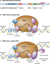
Fig 1
Fig 1
Interactions between holo RNA polymerase and promoters leading to transcript initiation. Panel (A) shows the key promoter sequence elements: each element is denoted by a colored rectangle positioned to indicate its location relative to position +1, the transcript start point. The sequence below each box denotes the consensus for E. coli σ70 holoenzyme. The labels −35, Ext, −10, Dis, and CRE denote the promoter −35 hexamer element, the extended −10 element, the −10 hexamer element, the discriminator element, and the core recognition element, respectively. Panel (B) illustrates the interaction of parts of the holo RNAP with different promoter elements in the closed complex. RNAP is drawn as a brown oval with the α subunit N- and C-terminal domains shown as blue circles. The four independently folding domains of the housekeeping σ subunit are shown as purple-shaded ovals marked σ1, σ2, σ3, and σ4, located to indicate the interactions described in the text. Panel (C) illustrates the interaction of parts of the holo RNAP with different promoter elements in the open complex. Using the same convention as in (B), the figure shows the transcription bubble with the template strand (orange) held by the CRE and the non-template strand (blue) held by σ Domain 2. (Adapted from reference .)
Fig 2
Fig 2
Regulation and roles of activatory transcription factors in bacteria. The left side of the figure notes that the function of an activatory factor (denoted by a yellow oval) can be regulated by (i) its level (set by its synthesis and/or its degradation), (ii) interaction with a protein partner, (iii) interaction with a ligand, (iv) sequestration to a location (e.g., the cytoplasmic face of the bacterial inner membrane), or (v) covalent modification. The right side of the figure illustrates how the active form of the factor may function by (i) replacing an RNAP subunit (e.g., the housekeeping σ subunit is replaced by an alternative σ), (ii) assisting RNAP to follow the “normal” pathway to transcript initiation, as in Fig. 1 (e.g., by promoting the interaction of αCTD with promoter DNA and σ), (iii) remodeling part of RNAP (e.g., by binding to the 265-determinant of αCTD, thereby changing its base sequence preferences), (iv) remodeling the promoter (e.g., by altering the juxtaposition between the promoter −10 and −35 elements), or (v) removing a repressor (shown as a red oval bound at a target promoter). In each case, specificity is determined by the recognition of target promoter sequence elements by the activatory factor. (Based on data from reference ; for simplicity, just one αCTD is shown.)
Fig 3
Fig 3
Activation by RNAP recruitment via αCTD. The upper line of the figure illustrates the two major strategies used by activators to recruit RNAP to target promoters via αCTD. The figure uses drawing conventions from Fig. 1 and 2, with functional interactions denoted by colored dots, listed in the inset box. Panel (A) illustrates assistance in which an activator–αCTD interaction promotes αCTD binding to promoter DNA and contact with σ Domain 4. Panel (B) illustrates remodeling, in which the activator contacts the DNA-binding 265-determinant of αCTD, thereby altering its binding specificity. The lower line illustrates three specific examples: in each case, specificity is determined by an operator sequence that is targeted by the activator. Panel (C) illustrates the activation of the E. coli lac operon promoter by CRP. An activating region (AR1) in the downstream subunit of the CRP dimer interacts with a determinant in αCTD (the 287-determinant), thereby promoting the interaction of the 265- and 261-determinants of αCTD with promoter DNA and σ Domain 4, respectively (59, 60). Here, for a productive interaction, CRP and αCTD must be bound to the same face of the DNA helix, and this can facilitate activation at other promoters by CRP bound at locations further upstream (61). Note that this type of activation is sometimes referred to as Class I activation (2). Panel (D) illustrates the activation of the ‘phage λpRE promoter by cII protein. An activating region in the upstream subunit of the cII tetramer interacts with the 271-determinant in αCTD, thereby promoting the interaction of the 265- and 261-determinants of αCTD with promoter DNA and σ Domain 4, respectively (62). Here, for a productive interaction, cII and αCTD must be bound to opposite faces of the DNA helix, and a similar arrangement is found at some promoters that are activated by members of the response-regulator family (63). Panel (E) illustrates the activation of the E. coli zwf promoter by SoxS that makes interactions with the DNA-binding 265-determinant of one αCTD. A second activating region contacts the other αCTD, thereby promoting its interaction with promoter DNA and σ Domain 4 (64). Note that other arrangements can be found at different SoxS-activated promoters (64, 65).
Fig 4
Fig 4
Activation by targeting Domain 4 of the RNAP σ subunit. The upper line of the figure illustrates the two major strategies used by activators that target RNAP σ4. Panel (A) illustrates assistance, in which an activator–σ4 interaction promotes σ Domain 4 binding to the promoter −35 element, and thereby open complex formation and transcript initiation: the activator binds to an operator sequence that abuts the promoter −35 element. Panel (B) illustrates remodeling, in which the activator contacts and repositions RNAP σ Domain 4. Essentially, the DNA-binding specificity of σ Domain 4 is complemented by, or replaced with, that of the activator protein. The figure uses the same drawing style as Fig. 3, and functional interactions are denoted by colored dots, listed in the inset box. The lower line illustrates three specific examples. Panel (C) illustrates the activation at ‘phage T4 middle promoters by the early phage-encoded AsiA and MotA proteins. Essentially, AsiA remodels host RNAP σ Domain 4 so that it becomes susceptible to activation by MotA (71–74). Panel (D) illustrates the activation of the E. coli micF promoter by SoxS that makes interactions with both the DNA-binding 265-determinant of one αCTD and with σ Domain 4 (64). The interactions, together, alter the binding preferences for RNAP in and upstream of the −35 region. Panel (E) illustrates the activation of the E. coli gal operon P1 promoter by CRP: note that galP1 is typical of the many E. coli promoters where CRP binds to a target that abuts the promoter −35 region (75). Here, αCTD is displaced and binds upstream, making a productive interaction with an activating region (AR1) in the upstream subunit of the CRP dimer. A second activating region (AR2) in the downstream CRP subunit interacts with a determinant in αNTD, while a third activating region (AR3) in the downstream CRP can contact σ Domain 4 (61, 76). Note that this type of activation is sometimes referred to as Class II activation (2). During Class II CRP-dependent activation, AR2 is the predominant activating region while, for other CRP family members, such as FNR, AR3 is predominant (61, 77, 78), and different adhesive activator–RNAP interactions can stabilize different intermediates along the pathway to transcript initiation (78, 79).
Fig 5
Fig 5
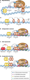
Mechanisms of promoter co-dependence on two activatory factors. The figure shows illustrations of each of the known mechanisms whereby the activity of a bacterial promoter can be dependent on two activators, shown as rust-colored and yellow ovals, denoted Activator 1 and Activator 2. The figure uses the same drawing style as Fig. 3 and 4, with some functional interactions denoted by colored dots, listed in the inset box. Panel (A) illustrates mechanisms in which Activator 2 is needed to position Activator 1 at a location where it is functional for activation. (i) illustrates activation with RNAP σ54 holoenzyme. The atypical σ54 is illustrated as a series of tangerine-shaded ovals, labeled according to reference (104). RNAP σ54 holo enzyme contacts promoter −24 and −12 elements (pink rectangles) but is unable to proceed from the closed to open complex, as the activity of determinants in the ELH-HTH region is occluded by the RI domain. The ATP-driven action of an enhancer-binding protein (Activator 1) is required to relieve this blockage so that the transcription bubble can open (facilitated by ELH-HTH, see text). At some σ54-dependent promoters, Activator 2 is required to ensure the correct positioning of Activator 1, by bending the upstream DNA. Note that, in some cases, Activator 2 also assists with the initial recruitment of RNAP σ54 holoenzyme by interacting with αCTD (110, 111). (ii) illustrates the original report of repositioning (112), where the binding of Activator 2 repositions Activator 1 from a location where it is unable to activate transcription to a location where it is able to activate transcription (in this case, Activator 2 is CRP and Activator 1 is MalT). Panel (B) illustrates co-dependence in which Activator 1 and Activator 2 bind independently to their targets and make independent but complementary contacts with different parts of RNAP σ70 holoenzyme. In the majority of such cases, as illustrated here, one activator contacts σ Domain 4, while the other contacts one of the displaced αCTDs, but there are some promoters where both activators bind further upstream and only contact αCTD (113). Panel (C) illustrates co-dependence in which a repressor blocks the function of Activator 1, and the role of Activator 2 is to stop the action of the repressor. This mechanism was discovered at the E. coli nir operon promoter, where FNR-dependent activation is suppressed by the repressive action of two NAPs, IHF and Fis, but repression is countered by the binding of NarL (114, 115). Panel (D) illustrates co-dependence in which the binding of one factor requires binding of the other and vice versa. The scenario requires direct interaction between Activator 1 and Activator 2. While infrequent in E. coli and Salmonella, direct interactions between different transcription factors are being discovered in other bacterial clades (116–118). (Evolved from reference with permission from Elsevier.)

References

    1. Browning DF, Busby SJW. 2016. Local and global regulation of transcription initiation in bacteria. Nat Rev Microbiol 14:638–650. doi:10.1038/nrmicro.2016.103 - DOI - PubMed
    1. Lee DJ, Minchin SD, Busby SJW. 2012. Activating transcription in bacteria. Annu Rev Microbiol 66:125–152. doi:10.1146/annurev-micro-092611-150012 - DOI - PubMed
    1. Kompaniiets D, Wang D, Yang Y, Hu Y, Liu B. 2023. Structure and molecular mechanism of bacterial transcription activation. Trends Microbiol:S0966-842X(23)00290-1. doi:10.1016/j.tim.2023.10.001 - DOI - PubMed
    1. Busby SJW. 2019. Transcription activation in bacteria: ancient and modern. Microbiology (Reading) 165:386–395. doi:10.1099/mic.0.000783 - DOI - PubMed
    1. Sutherland C, Murakami KS. 2018. An introduction to the structure and function of the catalytic core enzyme of Escherichia coli RNA polymerase. EcoSal Plus 8. doi:10.1128/ecosalplus.ESP-0004-2018 - DOI - PMC - PubMed

MeSH terms

LinkOut - more resources