. 2024 Mar 19;98(3):e0146923.
doi: 10.1128/jvi.01469-23. Epub 2024 Feb 12.

GCRV-II invades monocytes/macrophages and induces macrophage polarization and apoptosis in tissues to facilitate viral replication and dissemination

Affiliations

GCRV-II invades monocytes/macrophages and induces macrophage polarization and apoptosis in tissues to facilitate viral replication and dissemination

Ning Xia et al. J Virol. .

Abstract

Grass carp reovirus (GCRV), particularly the highly prevalent type II GCRV (GCRV-II), causes huge losses in the aquaculture industry. However, little is known about the mechanisms by which GCRV-II invades grass carp and further disseminates among tissues. In the present study, monocytes/macrophages (Mo/Mφs) were isolated from the peripheral blood of grass carp and infected with GCRV-II. The results of indirect immunofluorescent microscopy, transmission electron microscopy, real-time quantitative RT-PCR (qRT-PCR), western blot (WB), and flow cytometry analysis collectively demonstrated that GCRV-II invaded Mo/Mφs and replicated in them. Additionally, we observed that GCRV-II induced different types (M1 and M2) of polarization of Mo/Mφs in multiple tissues, especially in the brain, head kidney, and intestine. To assess the impact of different types of polarization on GCRV-II replication, we recombinantly expressed and purified the intact cytokines CiIFN-γ2, CiIL-4/13A, and CiIL-4/13B and successfully induced M1 and M2 type polarization of macrophages using these cytokines through in vitro experiments. qRT-PCR, WB, and flow cytometry analyses showed that M2 macrophages had higher susceptibility to GCRV-II infection than other types of Mo/Mφs. In addition, we found GCRV-II induced apoptosis of Mo/Mφs to facilitate virus replication and dissemination and also detected the presence of GCRV-II virus in plasma. Collectively, our findings indicated that GCRV-II could invade immune cells Mo/Mφs and induce apoptosis and polarization of Mo/Mφs for efficient infection and dissemination, emphasizing the crucial role of Mo/Mφs as a vector for GCRV-II infection.IMPORTANCEType II grass carp reovirus (GCRV) is a prevalent viral strain and causes huge losses in aquaculture. However, the related dissemination pathway and mechanism remain largely unclear. Here, our study focused on phagocytic immune cells, monocytes/macrophages (Mo/Mφs) in blood and tissues, and explored whether GCRV-II can invade Mo/Mφs and replicate and disseminate via Mo/Mφs with their differentiated type M1 and M2 macrophages. Our findings demonstrated that GCRV-II infected Mo/Mφs and replicated in them. Furthermore, GCRV-II infection induces an increased number of M1 and M2 macrophages in grass carp tissues and a higher viral load in M2 macrophages. Furthermore, GCRV-II induced Mo/Mφs apoptosis to release viruses, eventually infecting more cells. Our study identified Mo/Mφs as crucial components in the pathway of GCRV-II dissemination and provides a solid foundation for the development of treatment strategies for GCRV-II infection.

Keywords: GCRV-II; apoptosis; grass carp (Ctenopharyngodon idella); monocytes/macrophages; polarization; viral dissemination.


Conflict of interest statement

The authors declare no conflict of interest.

Figures
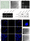
Fig 1
Fig 1
Mo/Mφ infection by GCRV-II in grass carp. (A) Morphology of Mo/Mφs after Giemsa staining under light microscopy. Scale bar = 20 µm. (B) Morphology of Mo/Mφs observed by TEM. N, nucleus; Scale bar = 2 μm. (C) Identification of cell purity. The specific marker genes were detected by RT-PCR in PBLs, Mo/Mφs, and no template control (N.T.C.). (D and E) Viral RNAs and protein determined by RT-PCR (D) and WB (E) after 48-h challenge of CIK cells and Mo/Mφs with GCRV-097 (MOI = 1). β-actin was employed as an internal control. (F) Subcellular localization of GCRV-II in CIK cells and Mo/Mφs under confocal microscopy. CIK cells and Mo/Mφs were challenged with GCRV-097 (MOI = 1) for 48 h. Blue represents the nucleus, and green indicates GCRV-II. (G) Mo/Mφs at indicated time points post-GCRV-097 challenge (MOI = 1) observed by TEM. The arrows point at the rupture of the cytomembrane. Scale bar = 2 μm.
Fig 2
Fig 2
TEM observation of CIK cells and Mo/Mφs post-GCRV-097 (MOI = 1) challenge for the indicated time. N, nucleus; LD, lipid droplets; and GCRV, GCRV-II particles. (A) TEM observation of mock Mo/Mφs. Scale bar = 2 μm. (B) GCRV-II-infected CIK cells under TEM. Scale bar = 200 nm. (C and D) GCRV-infected Mo/Mφs for 24 h (C) or 48 h (D) under TEM. Scale bar = 2 μm. The right panel is the magnification of TEM images. Scale bar = 200 nm.
Fig 3
Fig 3
GCRV-II replication in Mo/Mφs. (A and B) Relative mRNA expression levels of GCRV-II VP4 (A) and VP56 (B) in Mo/Mφs after challenge with GCRV-097 (MOI = 1) for the indicated time, determined by qRT-PCR. The relative mRNA expression level was normalized to that of the EF1a gene (n = 3). (C) Expression of GCRV-II VP4 protein in Mo/Mφs after challenge with GCRV-097 (MOI = 1) for the indicated time, determined by WB. (D–F) Percentages of GCRV-II-positive cells, determined by flow cytometry assay. (D) Mouse IgG group was used as a negative control. (E) Percentages of GCRV-II-positive cells in CIK cells after 60-h challenge with GCRV-097 (MOI = 1). (F) Percentages of GCRV-II-positive cells in Mo/Mφs after challenge with GCRV-097 (MOI = 1) for the indicated time. (G) Percentages of GCRV-II-positive cells from three separate experiments (*P < 0.05 and **P < 0.01).
Fig 4
Fig 4
GCRV-II-induced tissue macrophage polarization. (A) Relative mRNA expression levels of iNOS and arginase-2, the marker genes for M1 and M2 macrophages, respectively, in different tissues after 60-h challenge with GCRV-097, determined by qRT-PCR (n = 6). (B) Relative mRNA expression levels of IFN-γ2, IL-4/13A, and IL-4/13B, the marker genes inducing polarization toward M1 (IFN-γ2) and M2 (IL-4/13A and IL-4/13B), respectively, in different tissues after being challenged with GCRV-097 for 60 h, determined by qRT-PCR. The mRNA relative expression level was normalized to that of the 18s rRNA gene (n = 6). (C) Antibody specificity validation of iNOS (131 kDa) and arginase-2 (39 kDa). (D and E) Expression of iNOS and arginase-2 proteins in head kidney macrophages (D) and peripheral blood Mo/Mφs (E), determined by WB. The Mo/Mφs were isolated from the head kidney and peripheral blood of grass carp at 60 h after injection with PBS or GCRV-097 (1 × 10⁶ TCID50/mL, 4 μL/g). (F and G) Expression levels of iNOS and arginase-2 proteins in head kidney macrophages (F) and blood Mo/Mφs (G), determined by flow cytometry assay. (H and I) Relative fluorescence intensity of iNOS and arginase-2 in brain Mo/Mφs (H) and intestine Mo/Mφs (I), measured by ImageJ software, corresponding to the results of immunohistochemistry in Fig. S2. Brain and intestine were collected from the infected and control groups (n = 3) (*P < 0.05 and **P < 0.01).
Fig 5
Fig 5
Polarization of M1 and M2 macrophages in vitro. (A) SDS-PAGE analysis of purified recombinant intact CiIFN-γ2 (17.6 kDa), CiIL-4/13A (17.2 kDa), and CiIL-4/13B (17.1 kDa) proteins. (B and C) Measurement of CiIFN-γ2, CiIL-4/13A, and CiIL-4/13B protein activity by determining the phosphorylation levels of STAT5, ERK, and mTOR. (C) Measurement of phosphorylation levels of STAT5, ERK, and mTOR in three separate experiments by determining the gray value of the WB bands of CiIFN-γ2, CiIL-4/13A, and CiIL-4/13B proteins using the ImageJ software. (D and E) Verification of M1 macrophage polarization by mRNA expression levels of IL-1β, CXCR 3.1, and iNOS (D), as well as enzyme activity of iNOS (E). (F and G) Verification of M2 macrophage polarization by mRNA expression levels of IL-10, CXCR 3.2, and arginase-2 (F), as well as arginase-2 enzyme activity (G). Relative mRNA expression level was normalized to that of EF1a (n = 3) (*P < 0.05 and **P < 0.01).
Fig 6
Fig 6
High susceptibility of M2 macrophages to GCRV-II infection. (A and B) Relative mRNA expression levels of GCRV-II VP4 (A) and VP56 (B) in Mo/Mφs, M1, and M2 macrophages after being challenged with GCRV-097 (MOI = 1) for the indicated time, determined by qRT-PCR. The relative mRNA expression level was normalized to that of the EF1a gene (n = 3) (*P < 0.05 and **P < 0.01). (C) Expression levels of GCRV-II VP4 protein in Mo/Mφs, M1, and M2 macrophages after being challenged with GCRV-097 (MOI = 1) for 48 h, determined by WB. (D) Percentage of GCRV-II-positive cells in Mo/Mφs, M1, and M2 macrophages after being challenged with GCRV-097 (MOI = 1) for 24 and 48 h, determined by flow cytometry assay. Non-specific fluorescence was eliminated through mouse IgG. (E) Percentage of GCRV-II-positive cells from three separate experiments.
Fig 7
Fig 7
GCRV-II induces Mo/Mφs apoptosis to enhance self-replication. (A) Apoptosis rate of mock and infected Mo/Mφs, determined by flow cytometry assay. (B) Apoptosis rate of Mo/Mφs in three independent experiments. (C) Bcl-2 protein expression levels in Mo/Mφs at 60-h GCRV-097 (MOI = 1) challenge, determined by WB. (D) mRNA expression levels of Caspase-2, Caspase-7, Bcl2, and Mcl-1 in Mo/Mφs at 60-h post-GCRV-097 challenge (MOI = 1), determined by qRT-PCR. The relative mRNA level was normalized to that of the EF1a gene (n = 3). (E) Hoechst 33258 staining of Mo/Mφs at 60-h post-GCRV-097 challenge (MOI = 1). Scale bar = 10 μm. White arrows point to typical apoptotic symptoms of chromatin condensation, and red arrows point to typical apoptotic symptoms of nuclear fragmentation. (F) Incubation of Mo/Mφs with BH3 hydrochloride (BH3.h) (agonist) or Ilexsaponin A (antagonist) for 3 h and subsequent infection with GCRV-II (MOI = 1) for 60 h. The apoptosis rates of treated Mo/Mφs were measured by flow cytometry. (G and H) GCRV-II replication in Mo/Mφs, determined by qRT-PCR (G) and WB (H). Mo/Mφs were incubated with equal volumes of PBS, 200 ng/mL BH3.h (agonist), or 200 ng/mL Ilexsaponin A (antagonist) for 3 h and then infected with GCRV-II (MOI = 1) for 60 h. The relative mRNA expression level was normalized to that of the EF1a gene (n = 3). (I) GCRV-II replication in the brain, head kidney, intestine, and blood from different treatment groups, determined by qRT-PCR. Grass carp were injected intraperitoneally with equal volume of PBS, 10 µg/g BH3.h (agonist), or 10 µg/g Ilexsaponin A (antagonist) for 3 h and then infected with GCRV-097 (1 × 10⁶ TCID50/mL, 4 µL/g) for 60 h. The relative mRNA expression level was normalized to that of the 18s rRNA gene (n = 6) (*P < 0.05 and **P < 0.01).

Similar articles

Cited by

References

    1. Wang Q, Zeng W, Liu C, Zhang C, Wang Y, Shi C, Wu S. 2012. Complete genome sequence of a reovirus isolated from grass carp, indicating different genotypes of GCRV in China. J Virol 86:12466. doi:10.1128/JVI.02333-12 - DOI - PMC - PubMed
    1. Rao Y, Su J. 2015. Insights into the antiviral immunity against grass carp (Ctenopharyngodon idella) reovirus (GCRV) in grass carp. J Immunol Res 2015:670437. doi:10.1155/2015/670437 - DOI - PMC - PubMed
    1. Hou G, Lv Z, Liu W, Xiong S, Zhang Q, Li C, Wang X, Hu L, Ding C, Song R, Wang H, Zhang Y-A, Xiao T, Li J, Ledgerwood E. 2023. An aquatic virus exploits the Il6-STAT3-HSP90 signaling axis to promote viral entry. PLoS Pathog 19:e1011320. doi:10.1371/journal.ppat.1011320 - DOI - PMC - PubMed
    1. Hou G, Zhang Q, Li C, Ding G, Hu L, Chen X, Lv Z, Fan Y, Zou J, Xiao T, Zhang Y-A, Li J. 2023. An aquareovirus exploits membrane-anchored HSP70 to promote viral entry. Microbiol Spectr 11:e0405522. doi:10.1128/spectrum.04055-22 - DOI - PMC - PubMed
    1. Pei C, Ke F, Chen Z-Y, Zhang Q-Y. 2014. Complete genome sequence and comparative analysis of grass carp reovirus strain 109 (GCReV-109) with other grass carp reovirus strains reveals no significant correlation with regional distribution. Arch Virol 159:2435–2440. doi:10.1007/s00705-014-2007-5 - DOI - PubMed